

 PDF
PDF ePub
ePub Citation
 Print
Print


INTRODUCTION
Hand injuries caused by human bites are seen relatively often in clinical practice1. The majority of patients with human bites visit the hospital a considerable time after the injury. Thus, even when there are no clear findings of infection at the time of injury, infection should be considered when treating such cases. When human bite injuries are overlooked and progress to pyogenic arthritis or osteomyelitis, they become difficult to treat and severe sequelae in the joints can occur. Herein, with a review of the literature, we report a case involving wide debridement for pyogenic arthritis of the metacarpophalangeal joint resulting from a human bite. Subsequently, the patient showed a cartilage defect and severe instability, and was treated using an external fixator, with good clinical and radiologic outcomes.
CASE REPORT
A 12-year old boy was admitted to other hospital with an open wound acquired from a bite during a fight with a friend. At the time of admission, human bite wounds were observed on the dorsal and volar sides of the right index finger at the metacarpophalangeal joint. Exploration of the wounds was performed on the same day and a rupture of the extensor indicis proprius (EIP) was observed. He was treated with tenorrhaphy and primary closure of the wounds. During the post-procedure followup, the patient's pain became gradually more severe, and local heat and redness developed at the lesion site. Incision and drainage were performed approximately 2 weeks after the initial procedure; however, there was no improvement and the patient was transferred to our hospital.
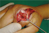
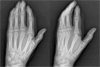
When the patient was admitted to our hospital, pus was flowing from where the wounds had been. Radiography revealed osteolysis and a loss of cartilage at the metacarpal head and base of the proximal phalanx, as well as subluxation of the metacarpophalangeal joint (Fig. 1). The patient was diagnosed with uncontrolled pyogenic arthritis and surgical treatment was performed. Surgical findings showed infection and degradation of the EIP tendon, radial collateral ligament, and dorsal capsule, as well as an articular cartilage defect (Fig. 2). After wide debridement and placement of a drainage catheter, the wound was closed with sutures and a splint fixation was performed.
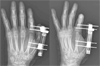
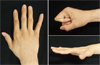
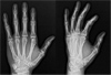
At 2 weeks postoperative, the infection was controlled. However, subluxation and severe instability of the radial side of the metacarpophalangeal joint persisted because of a metacarpal head defect and loss of the radial collateral ligament (Fig. 3). After applying gentle traction to correct the metacarpophalangeal joint, an external fixator was used to fix the metacarpophalgeal joint in a flexed position at 60° for 6 weeks (Fig. 4). The device was a commercialized product (Taeyeon Medical CO., Wonju, Korea) with a pin diameter of 2.3 mm. No bacterial grew in the intraoperative culture and cather tip culture. But we administered empirical intravenous second-generation cephalosporin for 3 weeks and intravenous aminoglycoside for 1 week. And then we continued to administer oral second-generation cephalosporin for 3 weeks. The patient was initially lost to follow-up after the removal of the external fixator. However, 6 years later, he visited the hospital in relation to a visa application; thus, a follow-up examination became possible. Although the metacarpophalangeal joint still showed a restricted range of motion of 0°–60° after 6 years, no instability was observed (Fig. 5). The joint had remodeled to resemble its normal shape, and the patient experienced no joint pain (Fig. 6). The patient currently plays basketball for his school team.
DISCUSSION
Pyogenic arthritis requires immediate treatment to avoid complications such as joint damage and osteomyelitis23. In the hand, pyogenic arthritis usually results directly from a penetrating joint injury, such as a bite injury. It can also be caused by pyogenic flexor tenosynovitis or an infected mucous cyst, if the infection spreads to the joint. When pyogenic arthritis is diagnosed, prompt treatment is required, including antibiotic medication and (following wound dressing in the surgical theater) drainage with thorough debridement. If clinical symptoms do not improve within 24–48 hours, repeated debridement is required2. Primary wound closure after a human bite can be very dangerous, as the underlying injury can be serious, even when the superficial wound appears innocuous. For example, serious complications occurred in the present case because this principle was not followed.
The prognosis for pyogenic arthritis of the hand is generally poor, mainly because the diagnosis is typically delayed. If a bite injury at the joint is not accompanied by a fracture, the pain may not be severe, which can result in the injury being ignoring for several days. Often the patient only visits the hospital when the infection has already started and the wound has become painful. In such cases, bacterial toxins and proteolytic enzymes are secreted, the articular cartilage is destroyed, and the adjacent bone can develop osteomyelitis. When patients are treated with debridement, the infected bone, cartilage, and soft tissue must be thoroughly removed; however, this can cause contracture or instability of the joint24. Moreover, even if the joint infection is well controlled, additional treatment is required in many cases, such as arthrodesis or, in severe cases, amputation. Giuffre et al.2 performed a retrospective analysis of 110 patients who had developed pyogenic arthritis of the hand, and reported that many patients did not visit the hospital on the day of the injury; instead, the patients visited the hospital an average of 5 days later, once symptoms of infection had already begun. Thirteen out of the 110 patients underwent arthrodesis and 14 patients underwent amputation because of uncontrolled infection or bone and soft tissue defects. The mean time from injury to first debridement was 3 months in the patients who underwent amputation, 20 days in the patients who underwent arthrodesis, and just 6 days in the patients who healed successfully after debridement. Thus, the prognosis deteriorates with increasing time until the first debridement.
Considerable debate exists regarding whether early joint mobilization is effective after surgery34567. Many authors report that early joint mobilization produces a good prognosis, similar to that for the treatment of pyogenic arthritis in other joints. However, Mennen and Howells8 reported that, following debridement, immobilization should be performed until symptoms of synovitis have disappeared. Some authors recommend fixation of the joint using a distraction external fixator after debridement. de Vries and van der Werken3 reported on the therapeutic outcomes of 20 patients with pyogenic arthritis of the hand; ultimately, 10 of the patients underwent arthrodesis or amputation and the overall outcomes were poor. However, 6 out of the 8 patients who wore an external fixator after the first debridement achieved almost complete functional recovery, suggesting that external fixation could become an important component in the treatment of pyogenic arthritis. Chadaev et al.6 also used an external fixator to distract the joint after debridement in 73 patients who showed cartilage and bone damage due to pyogenic arthritis of the metacarpophalangeal joint. Of the 73 patients, 39 (53%) showed a favorable outcome (a decrease of 15% or less in the range of motion), 24 (33%) showed a satisfactory outcome (a decrease of 15%–30% in the range of motion), and 10 (14%) patients showed an unsatisfactory outcome (a decrease of more than 30% in the range of motion). Furthermore, the use of an external fixator after debridement has been reported to prevent joint fusion.
In the present case, the patient developed pyogenic arthritis after a human bite injury, and although there was no delay in the patient seeing a physician, the infection may have developed because the initial debridement was insufficient. Because the dorsal approach of the metacarpophalangeal joint is difficult to access the volar side of the metacarpal head, a dorsal small incision at the time of initial debridement is likely to be cause of insufficient debridement. It is considered that debridement should be performed by exposing the volar part of the metacarpal head through an additional volar incision or a long incision on the dorsal side. A thorough debridement was performed after 2 weeks through long dorsal incision, and surgical findings showed a severe infection involving the articular cartilage, extensor tendon, articular capsule, and even the collateral ligament. The bone and soft tissue defects after the removal of the infected tissue resulted in severe instability, and the articular cartilage defect required fixation to be considered. However, because the patient was an adolescent who was still growing, and because some bone and cartilage regeneration could be expected, an external fixator was applied. As the scar tissue that developed from the infection could provide some compensation for the function of the injured ligament, the metacarpophalangeal joint was fixed in a flexed position. Although ligament reconstruction had not been performed, there was no instability due to the collateral ligament defect 6 years later, and plain radiography showed an almost complete regeneration of the bone and articular cartilage. The external fixator is thought to have restored the alignment of the metacarpophalangeal joint, which remained consistent during the healing period for the bone defect. This can be considered similar to the situation in Legg-Calvé-Perthes disease in infants, where containment of the femoral head within the acetabulum has a major impact on its regeneration. Nevertheless, the range of motion for the metacarpophalangeal joint was restricted to 0°–60°; the use of a hinged external fixator, as in Chadaev et al.6 might have resulted in a better range of motion.
It is important to determine whether the physeal growth arrest has occurred because he was a growing adolescent. No sequential radiographic follow-up was performed to determine whether there was any growth arrest. However, the ratio of the second metacarpal length to the third metacarpal length in the plain radiographs at the first visit was maintained at the final visit, suggesting that the growth arrest did not occur.
Along with a review of the literature, we reported our experience of a case of pyogenic arthritis at the metacarpophalangeal joint caused by a human bite, in which a good therapeutic outcome was achieved with thorough debridement and external fixation. In cases of pyogenic arthritis caused by human bite, the most important aspect of the treatment involves debridement and appropriate antibiotic use promptly after diagnosis. Physicians should be aware that a delayed diagnosis results in a worse prognosis. In addition, even when a delayed diagnosis leads to severe joint damage, the application of a distraction external fixator should be considered before arthrodesis or amputation in patients who are still growing. Even if the destruction of the joint is intense, we think that it is better not to do hasty arthrodesis or amputation, because of sufficient bony remodeling potential during adolescent.

 XML Download