

 PDF
PDF ePub
ePub Citation
 Print
Print


INTRODUCTION
Despite scaphoid fractures are primarily recognoized and treated appropriately, nonunion may occur in 5% to 15% of scaphoid fractures1. Scaphoid nonunion can be predisposed to premature carpal arthrosis and long-term disability23. And long-term follow-up after untreated scaphoid nonunion has shown radiological osteoarthritis4.
Various surgical methods have been presented for scaphoid nonunion. However, the ideal treatment for scaphoid nonunion remains uncertain567. Usually, bone graft and internal fixation are conventional treatment for scaphoid nonunion. Commonly used internal fixation devices include Kirschner (K) wires and compression screws8910. While compression screw can achieve a high predictability of bone union (80%–90%)7, some disadvantages were described in previous report. When scaphoid nonunion is treated with corticocancellous bone graft and screw fixation, there is difficulty in dealing with cavitary defects in estabilished scaphoid nonunions11. Beacuase compression screw devieces can further split the graft material or cause the graft to displace, it can result in malreduction, graft failure, and finally union failure.
Although compression screw showed more stable than K-wires fixation on biomechanical study12, several studies showed similar clinical results between compression screw and K-wires131415.
The purpose of this study was to evaluate the clinical and radiographic results of iliac bone grafting with K-wire fixation for treating scaphoid nonunion.
MATERIALS AND METHODS
Of 98 patients who underwent surgical treatment because of scaphoid nonunion between January 2007 and January 2016, 13 patients with scaphoid nonunion who were treated with iliac bone graft and K-wire fixation were ret-rospective reviewed. We usually use the iliac bone graft and compression screw fixation for scaphoid nonunion. When the nonunion site showed large cavitary defect or failure after screw fixation, we considered the use of K-wire fixation. Medical records of patients including gender, age, symptoms, follow up duration, and operative and radiological data were reviewed. The inclusion criteria were: (1) a nonunion of the scaphoid with duration of at least six months, (2) patient with followed up of more than 12 months. All patients had plain radiographs, preoperative computed tomography (CT) scans, and postoperative CT scans. The exclusion criteria were: (1) patient treated with compression screw or combination of compression screw and K-wire, (2) patient that treated with vascularized bone graft because of avascular necrosis.
Functional evaluation was performed using the modified Mayo wrist scoring system. Range of motion (ROM), grip strength, and pain were measured. Range of motion was measured using goniometer. It was compared to that of the contralateral side. Maximal grip strength of the injured side was measured and reported as a percentage of maximal strength of the contralateral side. Pain scale was self-reported and graded using a questionnaire. In addition to satisfaction score, a medication of the Mayo Wrist Scoring Chart was used for functional assessment, allowing for a total of 100 points for four categories.
Measurements of both preoperative and postoperative radiographs and CT scans were comparatively reviewed. Assessments were made on posteroanterior (PA) views of neutral and deviation position and lateral views with the forearm in neutral rotation. Severity of collapse was measured based on intrascaphoid (IS) angle16 and scapholunate (SL) angle17 on CT. Fracture union was defined according to the criteria described by Dias18 and assessed at final follow-up from radiographs or with CT scans if the radiological appearances were inconclusive.
1. Surgical technique and postoperative management
Surgical procedure was carried out under general anesthesia. The scaphoid was approached through the volar aspect by a longitudinal incision along the radial border of the flexor carpi radialis. Longitudinal capsulotomy was done to expose the scaphoid. The fibrous nonunion of the scaphoid was debrided and the necrotic tissue was removed. The distal part of the scaphoid was curetted to obtain a vascularized bed. The scaphoid humpback deformity was realigned by insertion of a wedge-shape bone graft from the iliac crest. Two or three of 1.4 mm K-wire s were drilled from the scaphoid tubercle through the bone block and into the proximal pole. Reduction and the position of the pin were controlled with a fluoroscope. The wrist was immobilized for two weeks in a below-elbow thumb spica splint with the thumb in abduction, leaving the interphalangeal joint free. After that, short-arm thumb spica cast was worn for an additional of eight weeks. At postoperative 10 to 12 weeks, if radiographs looked satisfactory, then K-wire s were removed under local anesthesia in the theater. Patients were permitted to resume normal activities for daily living at 12 weeks post-operatively.
2. Statistical analysis
All statistical analysis was performed using IBM SPSS ver. 21.0 (IBM Corporation, Somers, NY, USA). Descriptive statistics were reported as mean and standard deviation (SD) for continuous variables and number (and percentage where possible) for discrete assessments. Comparisons between preoperative and postoperative outcomes were analyzed using paired t-test. A p-value of less than 0.05 was considered statistically significant.
RESULTS
There were 13 males with a mean age of 30.2 years (range, 13 to 57 years). The mean time from injury to operation was 22.2 months (range, 7 to 72 months). Mean follow-up duration was 15.6 months (range, 12 to 24 months).
Bony union was achieved in 11 (84.6%) of 13 patients. The mean preoperative ROM was 62.5° (SD, 11.5°) for extension and 72.5° (SD, 12.7°) for flexion. Postoperative ROM was improved to a mean of 64.0° (SD, 13.5°) for extension and 74.2° (SD, 15.7°) for flexion. However, there was no statistical difference between preoperative ROM and postoperative ROM. The modified Mayo wrist score was significantly (p<0.05) improved from 64.0 (SD, 8.3) preoperatively to 87.5 (SD, 9.7) postoperatively. Excellent results were obtained in 6 (46.2%) patients, good results were achieved for 5 (38.5%) patients, and fair results were obtained for 2 (15.3%) patients. The lateral intrascaphoid angle measured on follow-up CT was significantly improved from 39.5 (SD, 12.5) preoperatively to 27.2 (SD, 5.9) at the final follow up (normal, <35°). The scapholunate angle was also significantly improved from 66.0 (SD, 8.5) preoperatively to 55.1 (SD, 7.8) at final follow up (normal, <35°) (Table 1).
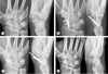
Complications occurred in six patients. Nonunion after treatment was occurred in two patients. Although they complain the pain and functional discomfort, they did not want to another operation and follow up was loss. In three patients, K-wire migration was found on radiograph. Although K-wire migration was shown, these patients obtained bony union of the nonunion site. After K-wire removal, there was no discomfort at the operation site (Fig. 1). One patient showed refracture after slip down.
DISCUSSION
The goal of surgical management of scaphoid nonunion is to improve wrist function. This includes bony union and restoring scaphoid alignment for functional improvement. It is known that re-establishment of the scaphoid length can relieves pain, improves grip strength and prevents degenerative changes17. To achieve these goals, a number of surgical approaches are available, including bone grafting, internal fixation, or a combination of both11.
Commonly used internal fixation devices include K-wire s and variable compression screw8910. K-wire s are easy to insert and remove. It can provide satisfactory stability. However, K-wire s are unable to provide compression at the fracture site. In addition, there is a need for extended postoperative immobilization which may lead to wrist stiffness. There are many different compression screws available to help achieve stable fixation when treating scaphoid nonunions. Usually, compression screws are cannluated, and headless screw. The cannulated aspect allows for easy insertion. Since the screw is headless, it may be buried under the articular surface. However, compression screw can further split the graft material or cause the graft to displace. Also, compression devices can cause the scaphoid shorten, which is undesirable for subsequent wrist mechanics.
Based on biomechanical stability study, compression screw can increase the strength of fixation and stiffness when compared to K-wires. Panchal et al.12 have studied the relative biomechanical stability of three types of internal fixation (Standard Acutrak screw, Mini-Acutrak screw, a pair of parallel 0.045-inch K-wires) with cancellous bone graft in a cadaveric scaphoid nonunion model. They concluded that Standard Acutrak screw might be a better option followed by the Mini-Acutrak screw and a pair of parallel 0.045-inch K-wire s when treating scaphoid nonunions. Although compression screw was more stable than K-wire s fixation in the biomechanical study, several clinical studies have shown similar results between compression screw and K-wire s with union rates ranging from 55% to 97%131415. The overall union rate of scaphoid nonunion treated by Herbert screw and bone grafting was 84%19. Our result (84.6%) are comparable with those of other studies of both K-wire and screw.
K-wire fixation technique has several advantages. K-wires occupy less of uniting surface area than a compression screw. This is a significant advantage in the context of difficult fracture healing seen in cavitary scaphoid nonunions. In addition, inadequately placed K-wires can be easily repositioned with minimal removal of bone. Moreover, two or three K-wires can increase the rotational stability on nonunion site when compared to one compression screw. Because of these advantages, K-wire fixation is considered a useful method for scaphoid nonunion. However, K-wire has also several disadvantage when we comparing with compression screw. It can be unable to provide compression at the fracture site. In addition, there is a need for extended postoperative immobilization which may lead to wrist stiffness.
This study has several limitations. First, the number of patients included in this study was small. In addition, the follow up period was relatively short. Moreover, we did not perform MRI to identify cases with avascular necrosis, which might affected the pathogenesis of nonunion and how to treat it. In addition, our study was a retrospective study. Nonetheless, this study revealed that iliac bone graft with K-wire fixation could provide good clinical and radiographic results for scaphoid nonunion.

 XML Download