Abstract
Brachial plexus injury is regarded as one of the most devastating injuries of the upper extremity. Accurate diagnosis is important to obtain the successful results. Basic preoperative evaluation includes simple radiography, cervical myelography. Magnetic resonance imaging, angiography, electrophysiologic studies and intraoperative studies. Furthermore, proper timing of surgery, surgical indication, plan and sufficient understanding of patients about the prognosis are the key for the satisfactory outcomes. This article provides an overview of the evaluation, diagnosis, intraoperative monitoring, and proper surgical planning for the treatment of posttraumatic brachial plexus injuries.
Go to : 
References
1. Bonney G. Prognosis in traction lesions of the brachial plexus. J Bone Joint Surg Br. 1959; 41:4–35.

3. Hentz VR, Narakas A. The results of microneurosurgical reconstruction in complete brachial plexus palsy: assessing outcome and predicting results. Orthop Clin North Am. 1988; 19:107–14.

4. Kawai H, Kawabata H, Masada K, et al. Nerve repairs for traumatic brachial plexus palsy with root avulsion. Clin Orthop Relat Res. 1988; (237):75–86.

5. Songcharoen P, Shin AY. Brachial plexus injury: acute diagnosis and treatment. Berger RA, Weiss AP, editors. Hand surgery. Philadelphia: Lippincott Williams & Wilkins;2004; 1005–25.
6. Limthongthang R, Bachoura A, Songcharoen P, Osterman AL. Adult brachial plexus injury: evaluation and management. Orthop Clin North Am. 2013; 44:591–603.
7. Robert EG, Happel LT, Kline DG. Intraoperative nerve action potential recordings: technical considerations, problems, and pitfalls. Neurosurgery. 2009; 65(4 Suppl):A97–104.
8. Kline DG. Timing for brachial plexus injury: a personal experience. Neurosurg Clin N Am. 2009; 20:24–6.

10. Hentz VR, Doi K. Traumatic brachial plexus injury. Wolfe S, Pederson W, Hotchkiss R, Kozin S, editors. Green’s operative hand surgery. Philadelphia: Elsevier;2010; 1235–94.
11. Mansat M. Surgical topographic anatomy of the brachial plexus. Rev Chir Orthop Reparatrice Appar Mot. 1977; 63:20–6.
12. Nagano A, Ochiai N, Sugioka H, Hara T, Tsuyama N. Usefulness of myelography in brachial plexus injuries. J Hand Surg Br. 1989; 14:59–64.

13. Doi K. Management of total paralysis of the brachial plexus by the double free-muscle transfer technique. J Hand Surg Eur Vol. 2008; 33:240–51.
15. Harper CM. Preoperative and intraoperative electrophysiologic assessment of brachial plexus injuries. Hand Clin. 2005; 21:39–46.

16. Kobayashi J, Mackinnon SE, Watanabe O, et al. The effect of duration of muscle denervation on functional recovery in the rat model. Muscle Nerve. 1997; 20:858–66.

17. Songcharoen P, Mahaisavariya B, Chotigavanich C. Spinal accessory neurotization for restoration of elbow flexion in avulsion injuries of the brachial plexus. J Hand Surg Am. 1996; 21:387–90.

18. Coulet B, Boretto JG, Lazerges C, Chammas M. A comparison of intercostal and partial ulnar nerve transfers in restoring elbow flexion following upper brachial plexus injury (C5-C6+/-C7). J Hand Surg Am. 2010; 35:1297–303.

19. Mackinnon SE, Novak CB, Myckatyn TM, Tung TH. Results of reinnervation of the biceps and brachialis muscles with a double fascicular transfer for elbow flexion. J Hand Surg Am. 2005; 30:978–85.

20. Liverneaux PA, Diaz LC, Beaulieu JY, Durand S, Oberlin C. Preliminary results of double nerve transfer to restore elbow flexion in upper type brachial plexus palsies. Plast Reconstr Surg. 2006; 117:915–9.

21. Chuang ML, Chuang DC, Lin IF, Vintch JR, Ker JJ, Tsao TC. Ventilation and exercise performance after phrenic nerve and multiple intercostal nerve transfers for avulsed brachial plexus injury. Chest. 2005; 128:3434–9.

Go to : 
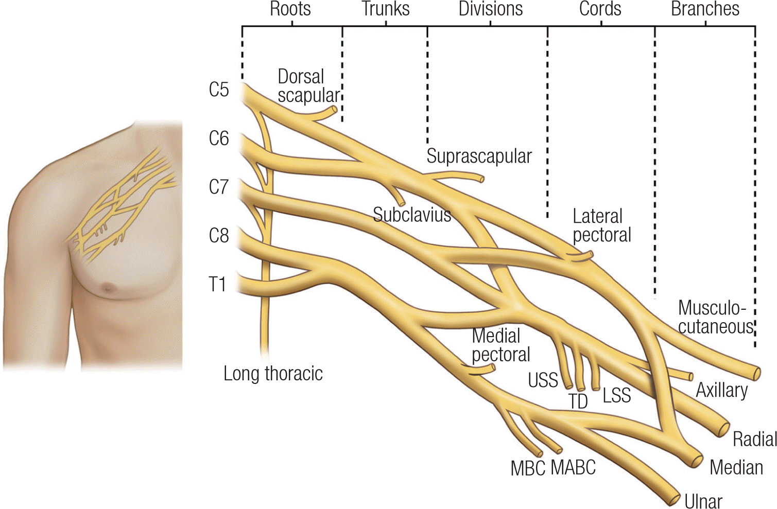 | Fig. 1.The anatomy of the brachial plexus. USS, upper subscapular; TD, thoracodorsal; LSS, lowere subscapular; MBC, medial brachial cutaneous; MABC, medial antebrachial cutaneus. |
 | Fig. 2.Traction injury of the brachial plexus. (A) Preganglionic injury cannot be repaired, (B) postganglionic stretch injury shows different magnitudes, and (C) extraforaminal rupture can be repaired with surgery |
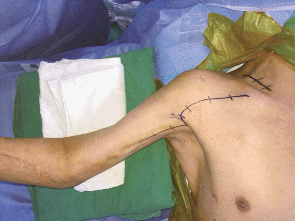 | Fig. 3.Double approach of brachial plexus injury (superior clavicular and deltopectoral approach). |
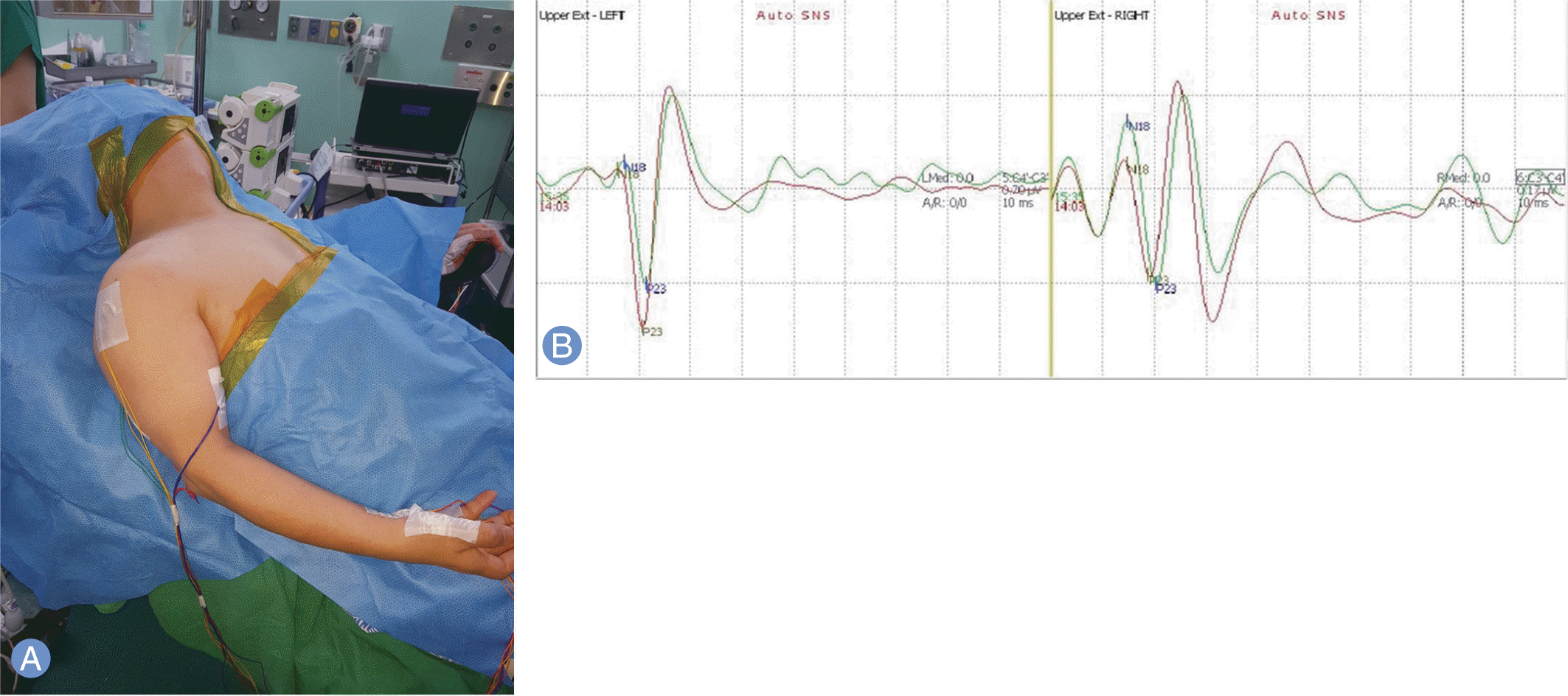 | Fig. 4.Electrophysiological evaluation during the surgery. (A) Intraoperative electro-physiologic monitoring of 55-year-old male with C8–T1 brachial plexus injury. (B) These lines means the preoperative and intraoperative base amplitude of C8-T1. SNS, sypathetic nerve stimultation. |
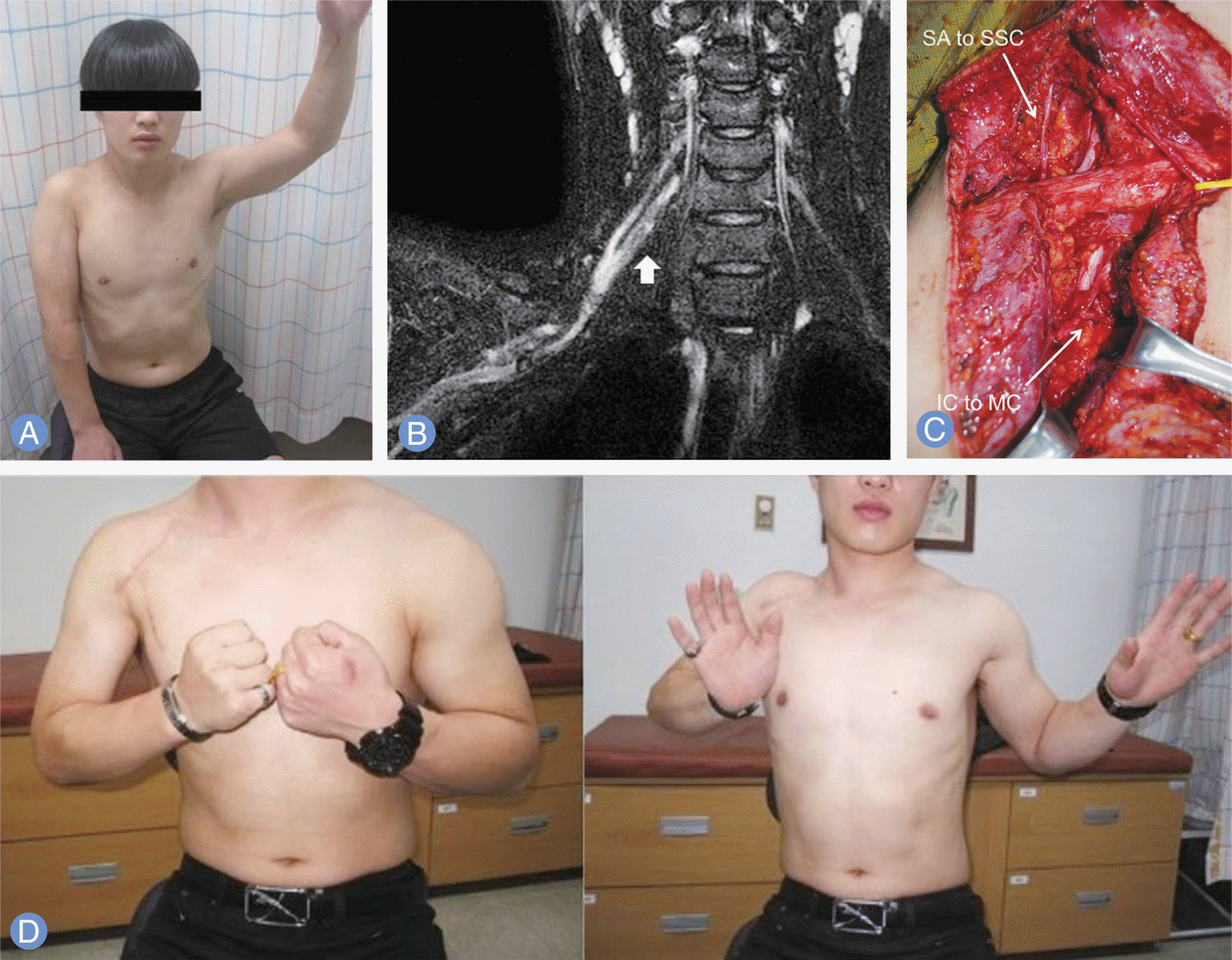 | Fig. 5.Nerve transfer of a 19 years-old male with right C5–6 brachial plexus injury after motorcycle accident. (A) The patient showed complete motor deficit in active elbow flexion and shoulder abduction. (B) Magnetic resonance image showed C5–6 nerve injury. (C) The 3–4 Intercostal nerves transfer to the musculocutaneous nerveand spinal accessory nerve transfer to the suprascapular nerve were performed at 7 months after injury. (D) Muscle strengths of elbow flexion and shoulder abduction were recovered at 16 months after surgery. SA, spinal accessory nerve; SSC, superior subscapular nerve; IC, intercostal nerve; MC, musculocutaneous nerve. |
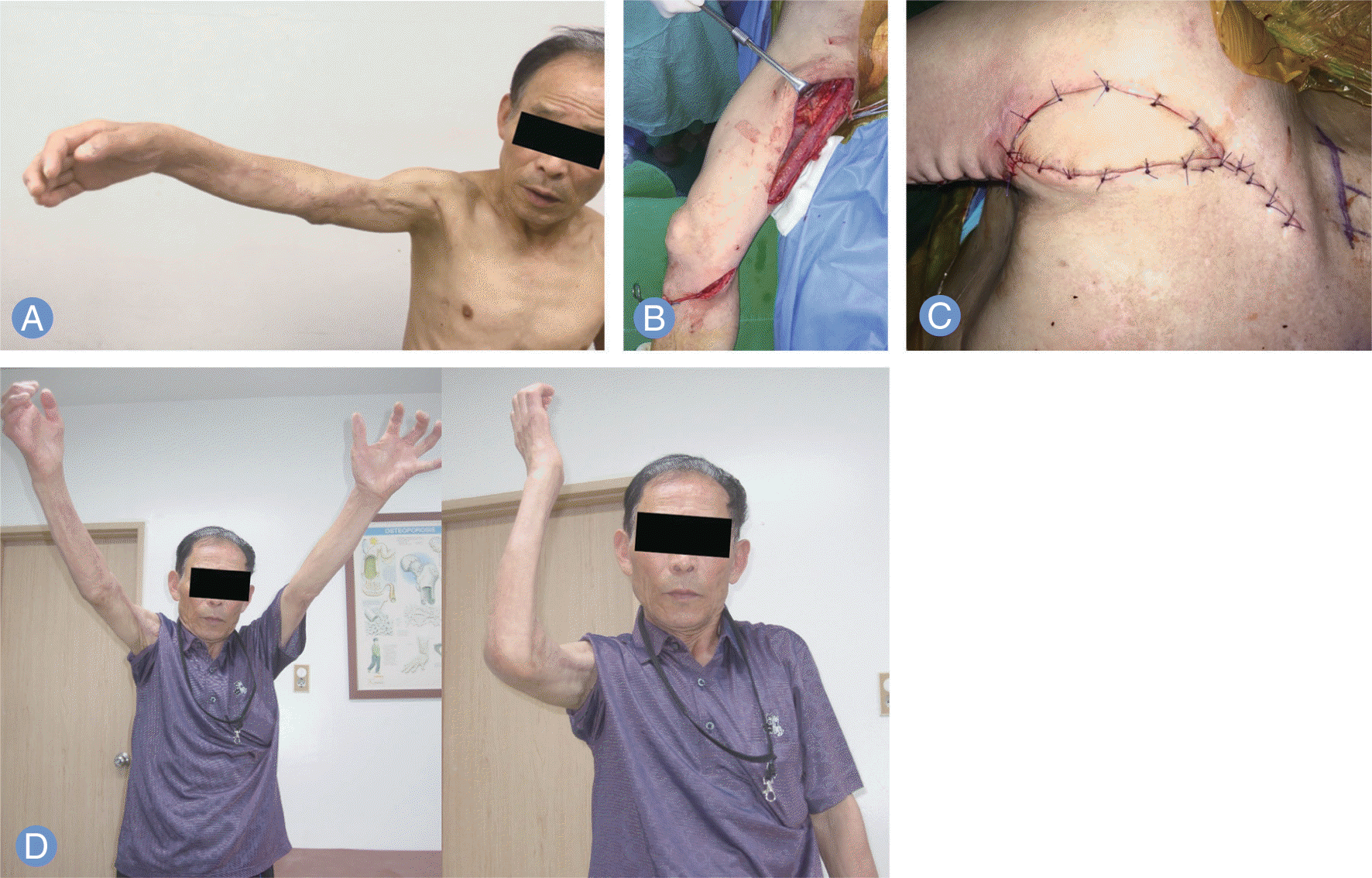 | Fig. 6.Functioning muscle transfer of a 64-year-old male who had crushing injury to his right arm at 1 year before the surgery. (A) The patient showed complete loss of active elbow flexion and shoulder abduction. (B) Gracilis muscle with monitoring flap was harvested from his right thigh. (C) Flap indicated well survival of free muscle transfer. (D) The patients showed excellent recovery of elbow flexion at 6 months after surgery. |
Table 1.
Clinical signs of preganglionic injury and its implications
Table 2.
Root innervation and motor functions in the upper extremity (from Robert et al.7)


 PDF
PDF ePub
ePub Citation
 Print
Print


 XML Download