Abstract
Purpose:
Out of nail components, nail plate, nail fold, paronychium, hyponychium, nail bed, and matrices are referred to as the perionychium. The authors report the outcomes of perionychial flap for reconstruction of fingertip injuries with nail bed injuries.
Methods:
We performed 8 cases of perionychial flap for fingertip injuries with nail bed injuries between January 2012 and December 2015, and analyzed the outcomes of the reconstruction surgery. The patients evaluated the aesthetic results on a four-point scale, and we measured and evaluated the ratio of axis length of the nail plate compared with collateral side of normal nail plate.
Results:
The mean follow up period was 8.4 months, and range of motion of distal interphalangeal joints and sensation of the reconstructed pulp were normal in all patients. After reconstructive surgery the nail plates regrew up to 80% in average compared to the normal side, and the satisfactory score were good to excellent as 3.8 point in average.
Go to : 
REFERENCES
1. Lee CH, Jeong JM, Kim J, et al. Nail lengthening using the eponychial flap. J Korean Orthop Assoc. 2009; 44:449–54.

2. Lee Y, Kwon S, Ko K. Correction of crooked nail deformity by modified osteoplastic reconstruction. Ann Plast Surg. 2000; 45:264–8.

4. Zook EG, Guy RJ, Russell RC. A study of nail bed injuries: causes, treatment, and prognosis. J Hand Surg Am. 1984; 9:247–52.

5. Bossley CJ. Conservative treatment of digit amputations. N Z Med J. 1975; 82:379–80.
6. Hosnuter M, Kargi E, Isikdemr A. An improvement in dorsal reverse adipofascial flap for fingertip reconstruction: nail matrix preservation. Ann Plast Surg. 2005; 55:155–9.
7. Tamai S. Twenty years’ experience of limb replantation: review of 293 upper extremity replants. J Hand Surg Am. 1982; 7:549–56.
10. Endo T, Nakayama Y. Microtransfers for nail and fingertip replacement. Hand Clin. 2002; 18:615–22.

11. Nakayama Y, Iino T, Uchida A, Kiyosawa T, Soeda S. Vascularized free nail grafts nourished by arterial inflow from the venous system. Plast Reconstr Surg. 1990; 85:239–45.

14. Brown RE, Zook EG, Russell RC. Fingertip reconstruction with flaps and nail bed grafts. J Hand Surg Am. 1999; 24:345–51.

15. Saito H, Suzuki Y, Fujino K, Tajima T. Free nail bed graft for treatment of nail bed injuries of the hand. J Hand Surg Am. 1983; 8:171–8.

16. Hsieh SC, Chen SL, Chen TM, Cheng TY, Wang HJ. Thin split-thickness toenail bed grafts for avulsed nail bed defects. Ann Plast Surg. 2004; 52:375–9.

17. Matev IB. Thumb reconstruction after amputation at the metacarpophalangeal joint by bone lengthening. J Bone Joint Surg Am. 1970; 52:957–65.
Go to : 
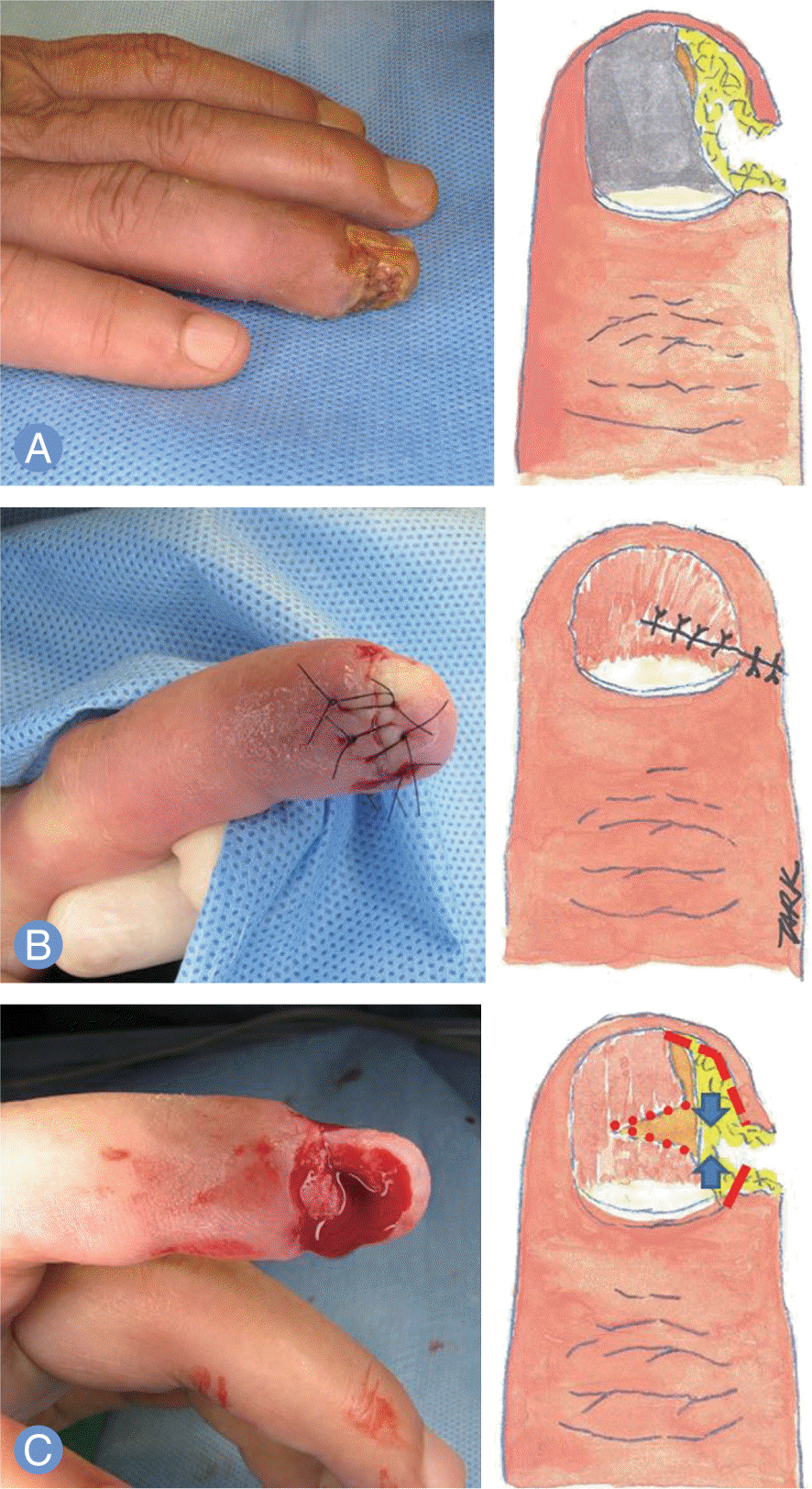
 | Fig. 1.Schematic design of perionychial flap for finger tip reconstruction. (A) Preoperative view. (B) After extraction of remaining nail, crushed soft tissues were minimally debrided. Comminuted fracture segments of the fingertip were also removed. Hyponychial, paronychial flap was elevated, and a triangular nail bed was excised to provide an ideal rotation of the hyponychial or paronychial flap. (C) Nail bed wounds were sutured with 5-0 Vicryl and 5-0 Nylon sutures. |
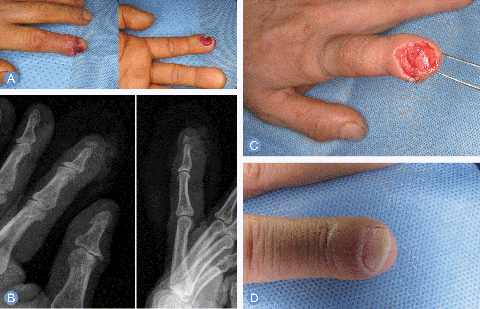
 | Fig. 2.
(A) A patient of 58-year-old man sustained a punch injury at a left index finger tip. (B) The radiographic image of the patient shows a fracture with a partial bone defect of the distal phalanx. (C) Reconstruction with a perionychial flap. (D) Postoperative 12 months view. |
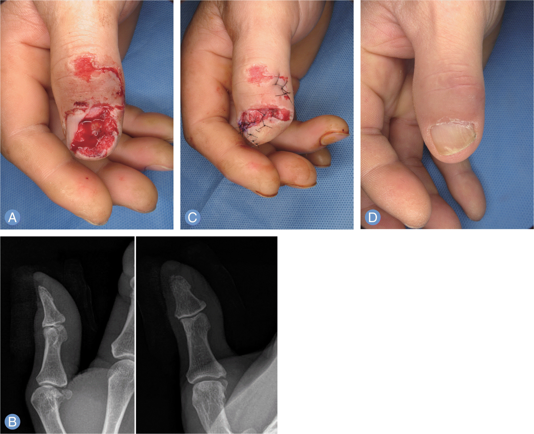
 | Fig. 3.
(A) A patient of 56-year-old man suffered from crushing injury at right thumb. (B) The radiographic image of the patient shows a comminuted fracture of the distal phalanx. (C) Reconstruction with a perionychial flap. (D) Postoperative 10 months view. |
Table 1.
Summary of the patients and postoperative follow-up


 PDF
PDF ePub
ePub Citation
 Print
Print


 XML Download