

 PDF
PDF ePub
ePub Citation
 Print
Print


Abstract
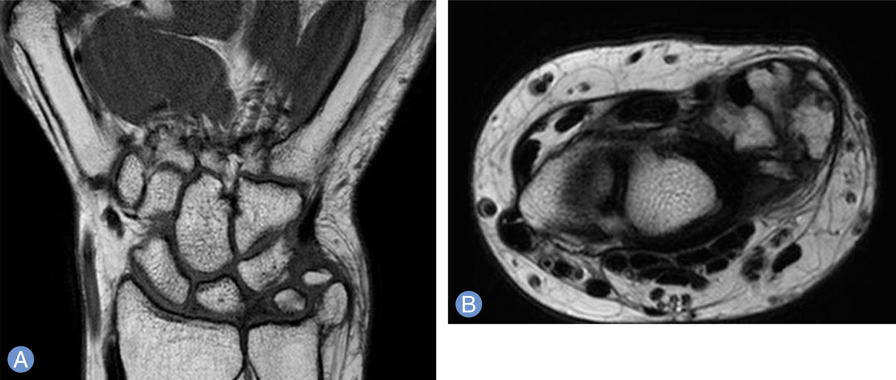
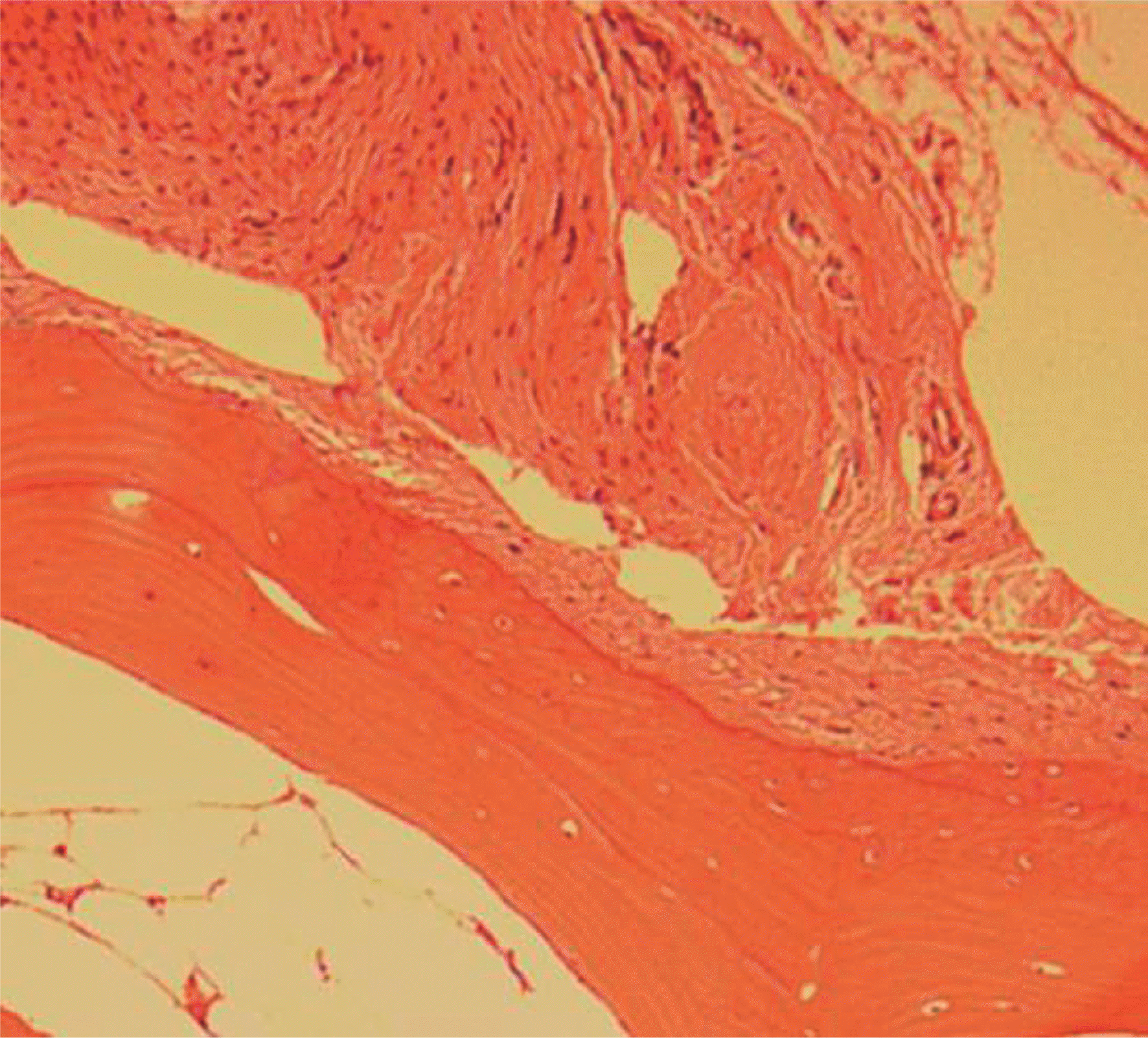
Synovial chondromatosis is a rare, benign and progressive metaplasia of the synovial membranes associated with the formation of cartilage in joints, tendon sheaths, or bursae. There are a few reports of synovial chondromatosis with wrist involvement. Here, we report a case of a 59-year-old woman with synovial chondromatosis of the ulnocarpal joint of the right wrist, with an 18-month follow-up and review of the literature.
REFERENCES
1. Ballet FL, Watson HK, Ryu J. Synovial chondromatosis of the distal radioulnar joint. J Hand Surg Am. 1984; 9:590–2.

2. Tudor A, Sestan B, Miletic D, et al. Synovial chondromatosis of the pisotriquetral joint with secondary osteoarthritis: case report. Coll Antropol. 2007; 31:1179–81.
3. Lee SK, Choy WS, Lee KW, Bae KJ. Synovial chondromatosis of the radiocarpal joint. Orthopedics. 2008; 31:811.
4. Inada Y, Fukui A, Maeda M, Tamai S, Inada M. Reconstruction of the triangular fibrocartilage complex after surgery for treatment of synovial osteochondromatosis of the distal radioulnar joint. J Hand Surg Am. 1990; 15:921–4.
5. Loonen MP, Schuurman AH. Recurrent synovial chondromatosis of the wrist: case report and literature review. Acta Orthop Belg. 2005; 71:230–5.
6. Wuisman PI, Noorda RJ, Jutte PC. Chondrosarcoma secondary to synovial chondromatosis: report of two cases and a review of the literature. Arch Orthop Trauma Surg. 1997; 116:307–11.
7. Burgess RC, Watson HK. Hypertrophic ulnar styloid nonunions. Clin Orthop Relat Res. 1988; (228):215–7.
8. Reverte Vinaixa MM, Singh R, Monyart JM, et al. Wrist synovial chondromatosis: case report and literature review. Hand Surg. 2012; 17:233–8.
9. Hallam P, Ashwood N, Cobb J, Fazal A, Heatley W. Malignant transformation in synovial chondromatosis of the knee? Knee. 2001; 8:239–42.
10. Constant E, Harebottle NH, Davis DG. Synovial chondromatosis of the hand: case report. Plast Reconstr Surg. 1974; 54:353–8.
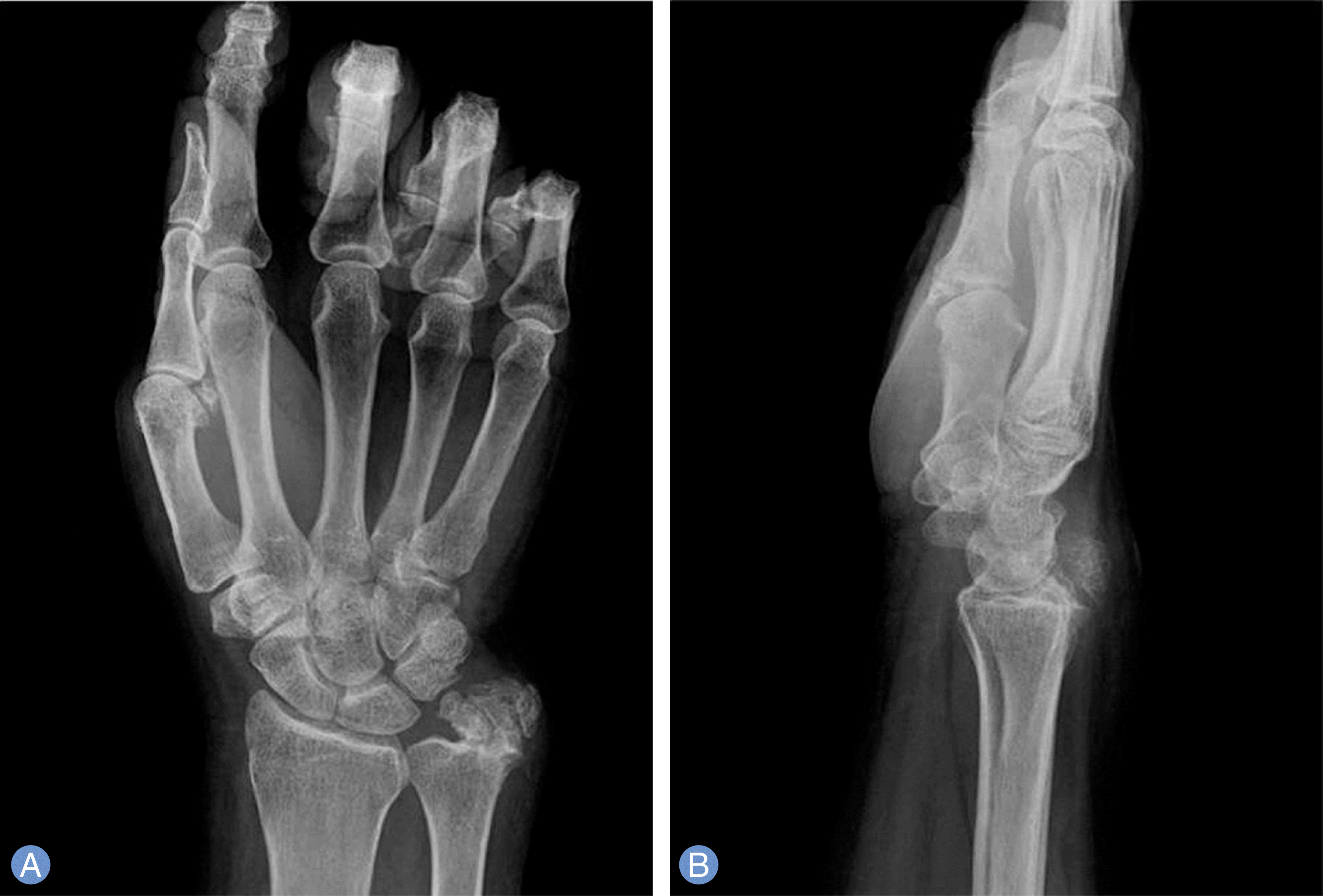
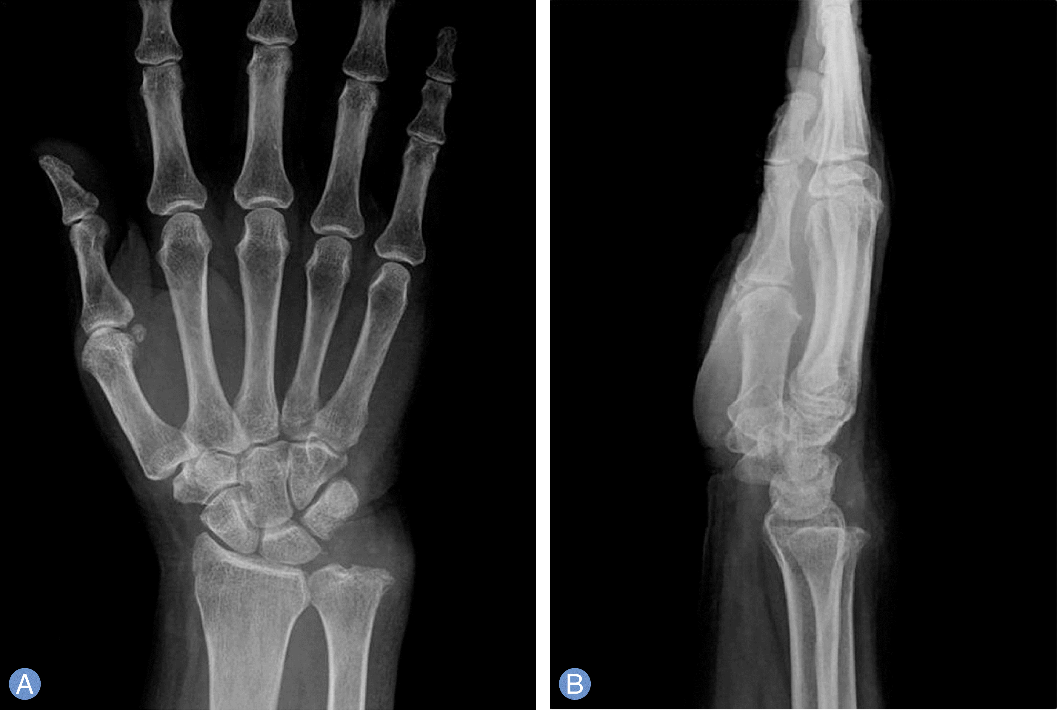
Fig. 1.
Multiple osseocartilaginous masses can be observed in the ulnocarpal joint on an anteroposterior radiograph (A) and in the dorsal aspect of the wrist on a lateral radiograph (B).
 XML Download