

 PDF
PDF ePub
ePub Citation
 Print
Print


Abstract
Posttraumatic instability of the carpometacarpal joint of the thumb are rarely reported. Consequently little is known about clinical and radiologic diagnosis, natural progress or treatment method. We report three cases of chronic instability of the carpometacarpal joint of the thumb treated with Eaton and Littler's ligament reconstruction. Satisfactory thumb functions were restored without arthritic change or recurrent subluxation.
REFERENCES
1. Eaton RG, Littler JW. Ligament reconstruction for the painful thumb carpometacarpal joint. J Bone Joint Surg Am. 1973; 55:1655–66.

2. Pellegrini VD Jr, Olcott CW, Hollenberg G. Contact patterns in the trapeziometacarpal joint: the role of the palmar beak ligament. J Hand Surg Am. 1993; 18:238–44.
3. Edmunds JO. Traumatic dislocations and instability of the trapeziometacarpal joint of the thumb. Hand Clin. 2006; 22:365–92.
4. Takwale VJ, Stanley JK, Shahane SA. Post-traumatic instability of the trapeziometacarpal joint of the thumb: diagnosis and the results of reconstruction of the beak ligament. J Bone Joint Surg Br. 2004; 86:541–5.
5. Park JS, Kim HK, Jung YK, Yoo JH, Kwon IH, Rah J. Ligament reconstruction for the posttraumatic instability of the carpometacarpal joint of the thumb: a report of three cases. J Korean Orthop Assoc. 2008; 43:112–7.
6. Simonian PT, Trumble TE. Traumatic dislocation of the thumb carpometacarpal joint: early ligamentous reconstruction versus closed reduction and pinning. J Hand Surg Am. 1996; 21:802–6.
7. Strauch RJ, Behrman MJ, Rosenwasser MP. Acute dislocation of the carpometacarpal joint of the thumb: an anatomic and cadaver study. J Hand Surg Am. 1994; 19:93–8.
8. Colman M, Mass DP, Draganich LF. Effects of the deep anterior oblique and dorsoradial ligaments on trapeziometacarpal joint stability. J Hand Surg Am. 2007; 32:310–7.
9. Bettinger PC, Linscheid RL, Berger RA, Cooney WP 3rd, An KN. An anatomic study of the stabilizing ligaments of the trapezium and trapeziometacarpal joint. J Hand Surg Am. 1999; 24:786–98.
10. Zhang X, Shao X, Huang W, Zhu H, Yu Y. An alternative technique for stabilisation of the carpometacarpal joint of the thumb after dislocation or subluxation. Bone Joint J. 2015; 97B:1533–8.
Fig. 1.
Intraoperative photographs for the reconstruction of the volar oblique ligament according to the Eaton and Littler technique. (A, B) Radial half of the flexor carpi radialis was passed through a drill hole in the base of the first metacarpal bone. (C) It was placed beneath the abductor pollicis longus then passed around the remaining flexor carpi radialis and secured over the dorsal capsule.
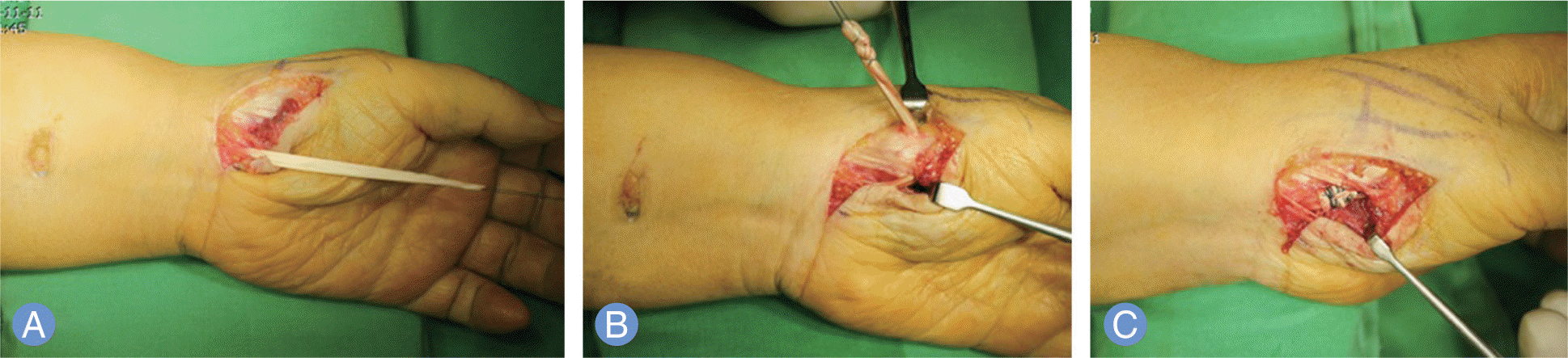
Fig. 2.
(A, B) Plain radiographs at 3 days after injury showed normal positioning of the carpometacarpal joint of the thumb with uniform joint space. (C) Antero-posterior radiograph of the wrist at 4 months after injury showed radially displaced carpometacarpal joint of the thumb. (D) Oblique radiograph of semipronated wrist at 4 months after injury showed widened interval between first and second metacarpal bone base and non-parallel joint space in comparison with initial radiograph.
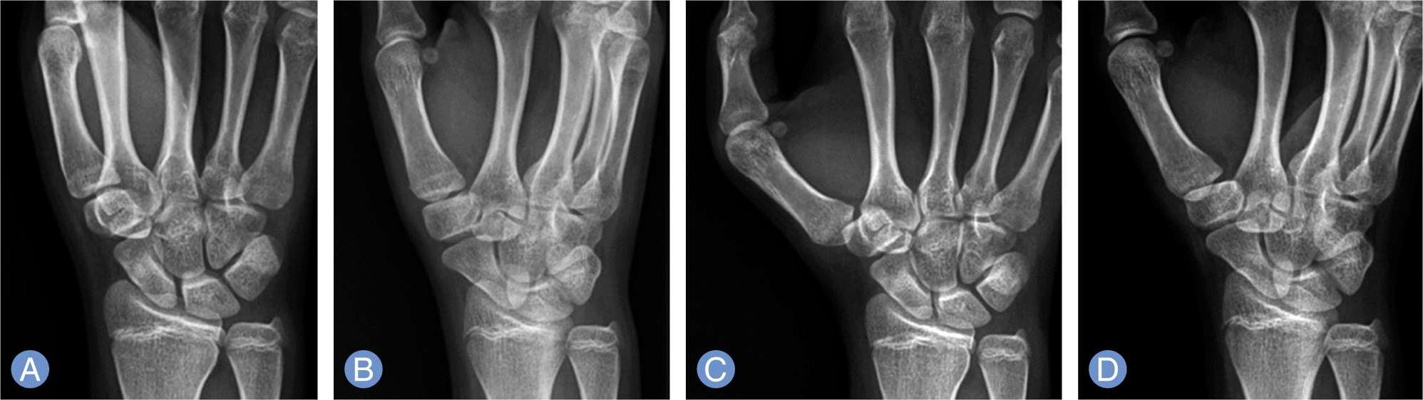
Fig. 3.
Two dimensional computed tomography coronal image suggested an air density (arrow) in the carpometacarpal joint of the thumb.
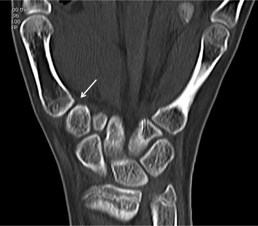
Fig. 4.
(A) Radiograph demonstrating reduced carpometacarpal joint of the thumb with a hole at the metacarpal base made to pass the split of flexor carpi radialis tendon. (B) Follow-up radiograph at 23 months after the surgery shows congruent joint with no arthritis.
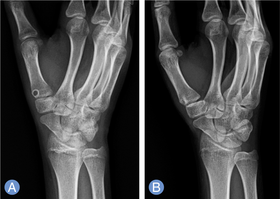
Fig. 5.
(A, B) Follow-up photograph at 23 months after the surgery shows full range of motion in all planes.
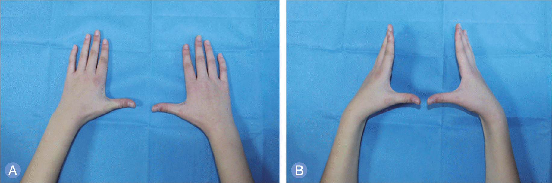
Fig. 6.
(A) Plain radiograph made at several days after injury in a local clinic showed normal alignment of the carpometacarpal joint of the thumb with uniform joint space. (B) Radiograph of the wrist at 1 year after injury showed radially displaced carpometacarpal joint of thumb. (C) Follow-up radiograph 18 months after the surgery showed sustained alignment with no arthritic change.
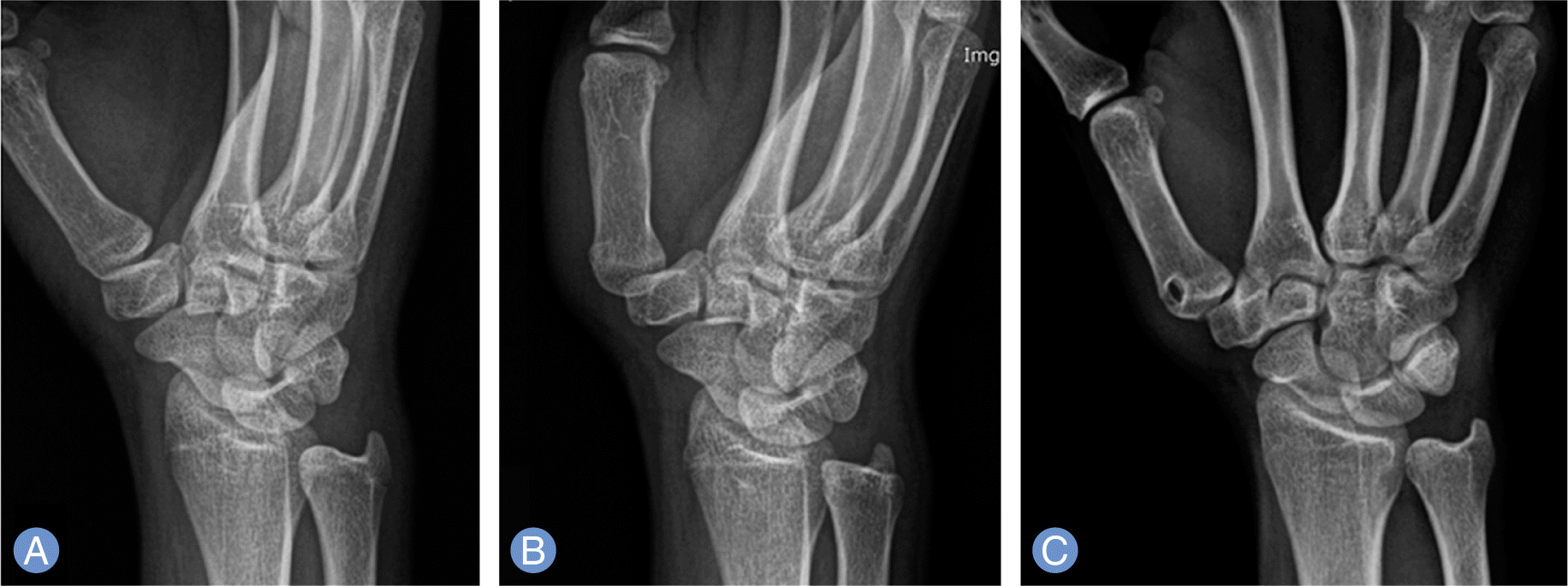
Fig. 7.
(A, B) Follow-up photograph at 18 months after the surgery shows full range of motion in all planes.
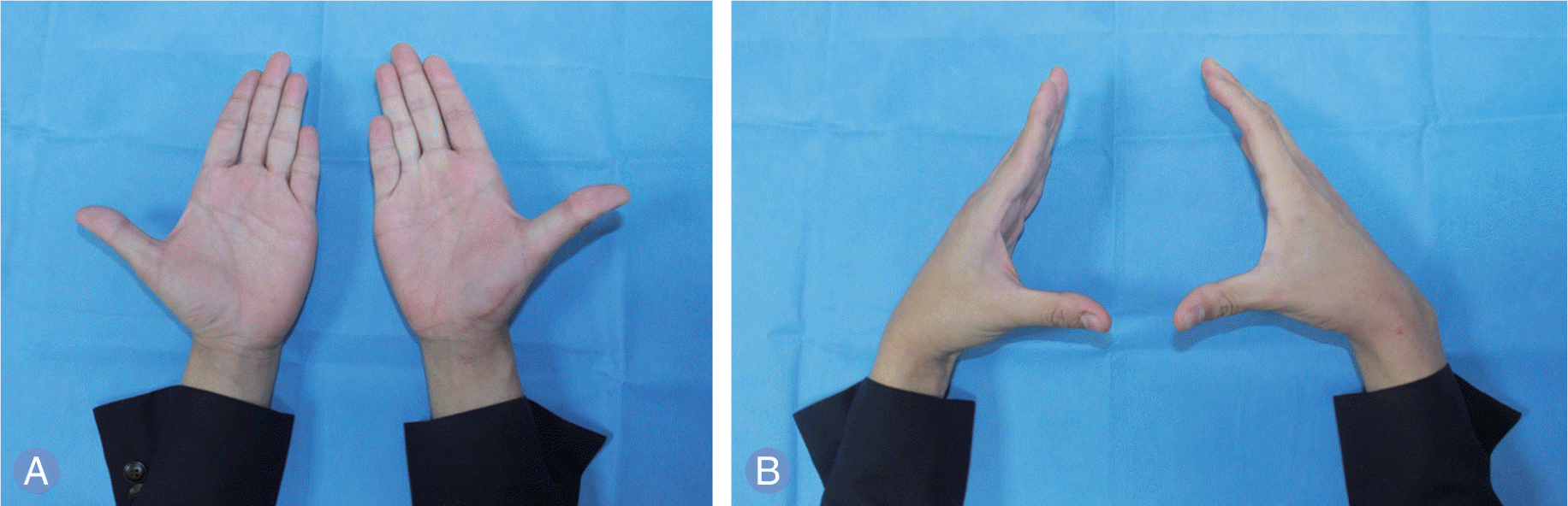
Fig. 8.
(A) Plain radiograph made after injury showed normal alignment of the carpometacarpal joint of the thumb with uniform joint space. (B) Radiograph of the wrist at 1 month after injury showed radially displaced carpometacarpal joint of the thumb. (C) Follow-up radiograph at 12 months after the surgery showed congruent joint space.
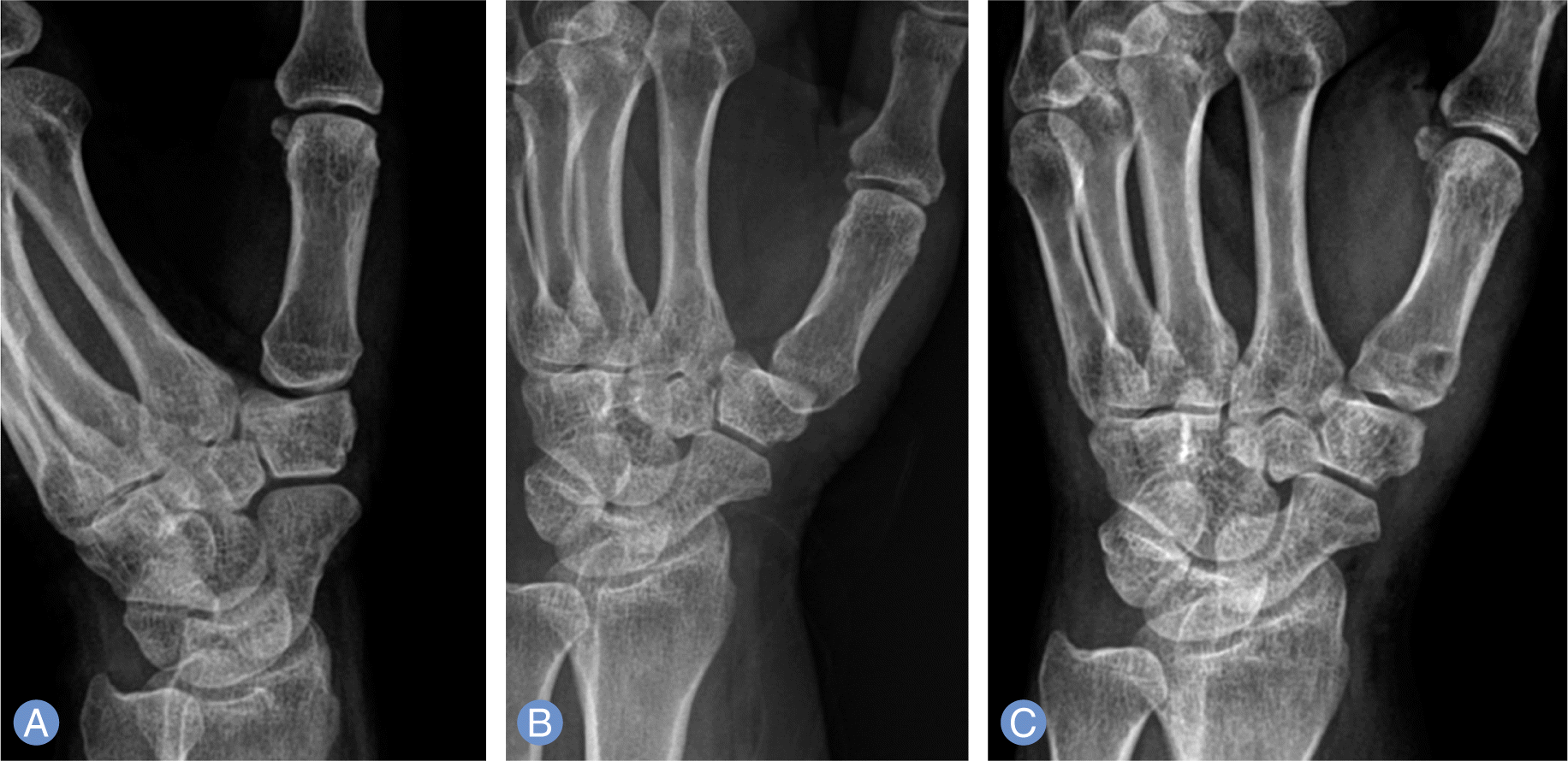
Fig. 9.
T2 weighted magnetic resonance coronal image (A) and T1 weighted sagittal image (B) suggested no specific abnormal finding at volar oblique ligament (arrows) in the carpometacarpal joint of the thumb.
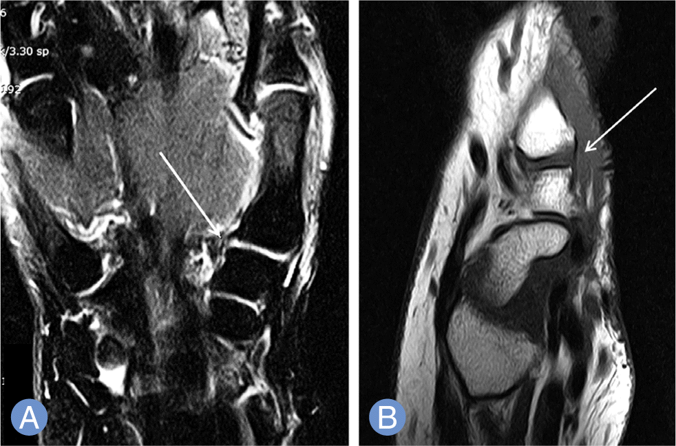
Table 1.
Summary of the case
 XML Download