Abstract
Purpose:
There remains uncertain whether to fix or not an ulnar styloid fracture acommpanied by distal radius fracture. Fixation might be required in cases of the fracture involving a fovea of ulnar head, an attachment site of deep triangular fibrocartilage, which is thought to be important to distal radioulnar joint stability. We analyzed a fovea involvement of an accompanied ulnar styloid fracture in patients with distal radius fracture by simple radiograph and three-dimensional computed tomography (3D CT).
Methods:
We retrospectively reviewed 168 patients who underwent surgery with volar locking plate for distal radius fracture in our hospital from January 2005 to March 2015 and evaluated a fovea involvement of ulnar head by simple radiographs and 3D CT respectively, and compared.
Results:
On simple X-ray, 64 cases (38%) were ulnar styloid fovea fractures; however, 21 cases of these revealed non-fovea fractures by 3D CT. And 7 out of 104 cases determined as non-fovea fracture by simple radiographs were diagnosed as fovea fractures by 3D CT. Sensitivity, specificity and accuracy of evaluation by simple radiograph were 86%, 82% and 83% respectively, when compared with those of 3D CT based evaluation.
Conclusion:
Accuracy of evaluating an accompanied ulnar styloid fovea fracture in patients with distal radius fracture by simple radiograph, when compared with 3D CT, was 83%; therefore, we recommend using the 3D CT based evaluation instead of simple radiograph based one for determination of fovea involvement of ulnar head.
Go to : 
REFERENCES
1. Frykman G. Fracture of the distal radius including sequelae: shoulder-hand-finger syndrome, disturbance in the distal radio-ulnar joint and impairment of nerve function: a clinical and experimental study. Acta Orthop Scand. 1967; 38(Suppl 108):31–61.
2. Oskarsson GV, Aaser P, Hjall A. Do we underestimate the predictive value of the ulnar styloid affection in Colles fractures? Arch Orthop Trauma Surg. 1997; 116:341–4.

3. Stoffelen D, De Smet L, Broos P. The importance of the distal radioulnar joint in distal radial fractures. J Hand Surg Br. 1998; 23:507–11.

4. May MM, Lawton JN, Blazar PE. Ulnar styloid fractures associated with distal radius fractures: incidence and implications for distal radioulnar joint instability. J Hand Surg Am. 2002; 27:965–71.

5. Kim JK, Koh YD, Do NH. Should an ulnar styloid fracture be fixed following volar plate fixation of a distal radial fracture? J Bone Joint Surg Am. 2010; 92:1–6.

6. Souer JS, Ring D, Matschke S, Audige L, Marent-Huber M, Jupiter JB. Effect of an unrepaired fracture of the ulnar styloid base on outcome after plate-and-screw fixation of a distal radial fracture. J Bone Joint Surg Am. 2009; 91:830–8.

7. Zenke Y, Sakai A, Oshige T, Moritani S, Nakamura T. Treatment with or without internal fixation for ulnar styloid base fractures accompanied by distal radius fractures fixed with volar locking plate. Hand Surg. 2012; 17:181–90.

8. Ozasa Y, Iba K, Oki G, Sonoda T, Yamashita T, Wada T. Nonunion of the ulnar styloid associated with distal radius malunion. J Hand Surg Am. 2013; 38:526–31.

9. Gogna P, Selhi HS, Mohindra M, Singla R, Thora A, Yamin M. Ulnar styloid fracture in distal radius fractures managed with volar locking plates: to fix or not? J Hand Microsurg. 2014; 6:53–8.

10. Yan YQ, Zhang PX, Wang TB, Chen JH, Jiang BG. Case-control study on effects of fracture of processus styloideus ulnae on prognosis after plate fixation for the treatment of distal radial fractures. Zhongguo Gu Shang. 2015; 28:226–9.
11. Yilmaz S, Cankaya D, Karakus D. Ulnar styloid fracture has no impact on the outcome but decreases supination strength after conservative treatment of distal radial fracture. J Hand Surg Eur Vol. 2015; 40:872–3.

12. Buijze GA, Ring D. Clinical impact of United versus nonunited fractures of the proximal half of the ulnar styloid following volar plate fixation of the distal radius. J Hand Surg Am. 2010; 35:223–7.

13. Hauck RM, Skahen J 3rd, Palmer AK. Classification and treatment of ulnar styloid nonunion. J Hand Surg Am. 1996; 21:418–22.

14. Sammer DM, Chung KC. Management of the distal radioulnar joint and ulnar styloid fracture. Hand Clin. 2012; 28:199–206.

15. Ishii S, Palmer AK, Werner FW, Short WH, Fortino MD. An anatomic study of the ligamentous structure of the triangular fibrocartilage complex. J Hand Surg Am. 1998; 23:977–85.

16. Shaw JA, Bruno A, Paul EM. Ulnar styloid fixation in the treatment of posttraumatic instability of the radioulnar joint: a biomechanical study with clinical correlation. J Hand Surg Am. 1990; 15:712–20.

17. Mirarchi AJ, Hoyen HA, Knutson J, Lewis S. Cadaveric biomechanical analysis of the distal radioulnar joint: influence of wrist isolation on accurate measurement and the effect of ulnar styloid fracture on stability. J Hand Surg Am. 2008; 33:683–90.

18. Nakamura R, Horii E, Imaeda T, Nakao E, Shionoya K, Kato H. Ulnar styloid malunion with dislocation of the distal radioulnar joint. J Hand Surg Br. 1998; 23:173–5.

19. Gong HS, Cho HE, Kim J, Kim MB, Lee YH, Baek GH. Surgical treatment of acute distal radioulnar joint instability associated with distal radius fractures. J Hand Surg Eur Vol. 2015; 40:783–9.

20. Yanagida H, Ishii S, Short WH, Werner FW, Weiner MM, Masaoka S. Radiologic evaluation of the ulnar styloid. J Hand Surg Am. 2002; 27:49–56.

21. van Der Heijden B, Groot S, Schuurman AH. Evaluation of ulnar styloid length. J Hand Surg Am. 2005; 30:954–9.

22. Karl JW, Swart E, Strauch RJ. Diagnosis of occult scaphoid fractures: a cost-effectiveness analysis. J Bone Joint Surg Am. 2015; 97:1860–8.
Go to : 
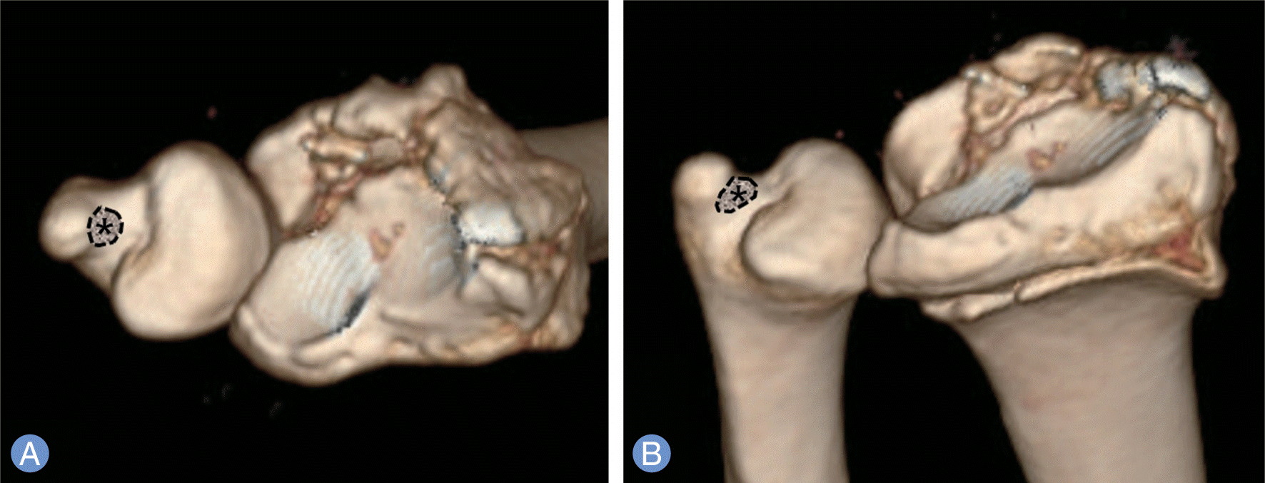
 | Fig. 1.Fovea of ulnar styloid process. (*, asterisk in dotted line). (A) Axial view of three-dimensional computed tomography (3D CT) reconstruction image. (B) Anteroposterior-oblique view of 3D CT reconstruction image. |
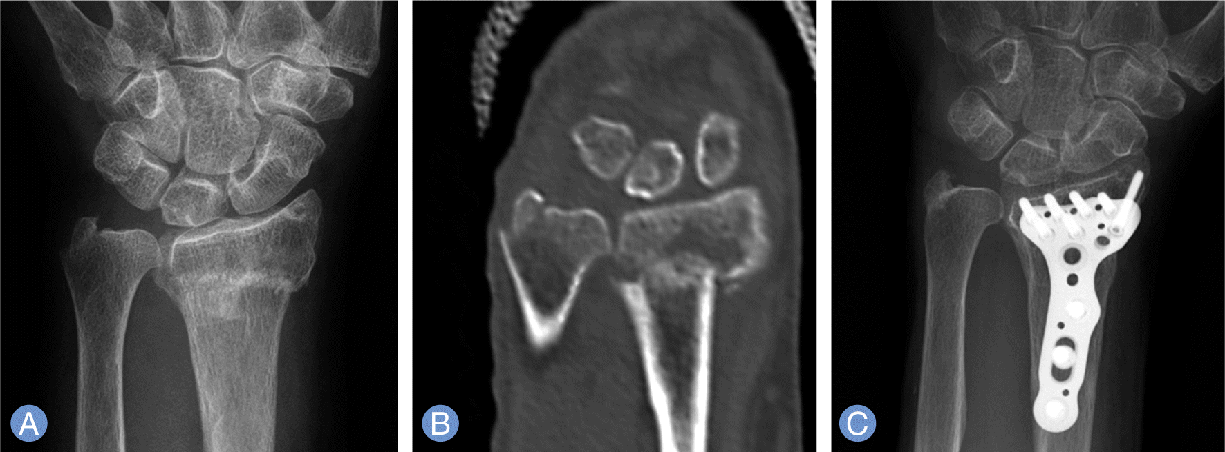
 | Fig. 2.Initial posteroanterior radiographs (A) of right wrist of 82-year-old female patient showed ulnar styloid base fracture. It seems like that there is a fovea involvement of fracture line on simple radiograph, but, considering computed tomography reconstruction centered by styloid process (B), fovea is not involved, and deep triangular fibrocartilage was supposed to be intact.). Radiograph taken at postoperative 6 months (C) shows a healed fracture of styloid process without surgery. |
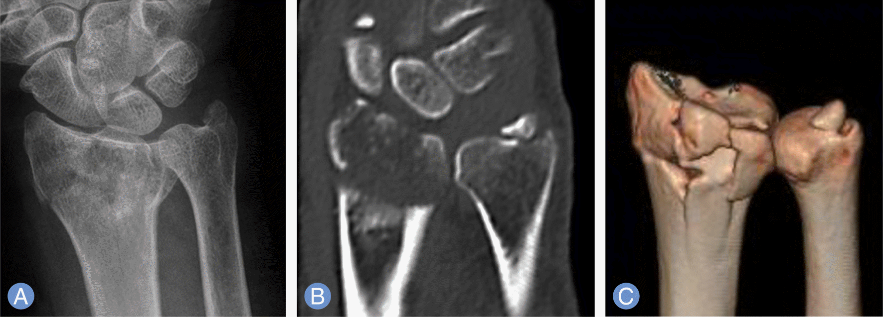
 | Fig. 3.Initial posteroanterior radiograph of left wrist of 61-year-old lady (A) was interpreted as non-base fracture of ulnar styloid, but the coronal (B) and three-dimensional (3D) reconstruction image (C) of computed tomography scan showed a fovea involvement. 3D reconstruction image (C) revealed more clear figuration of displaced styloid fragment with foveal beak. |
Table 1.
Distribution of ulnar styloid base and non-base fracture evaluated by simple radiograph and 3D CT


 PDF
PDF ePub
ePub Citation
 Print
Print


 XML Download