Abstract
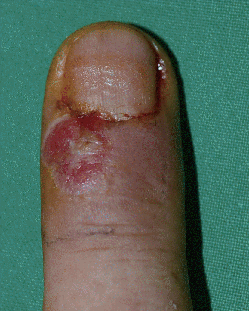
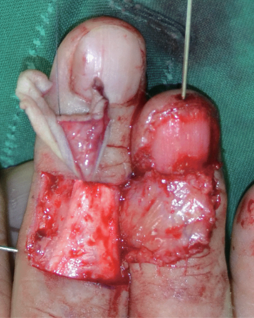
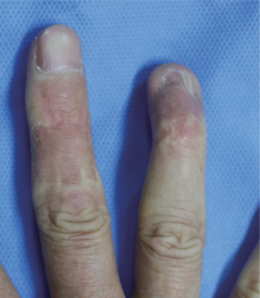
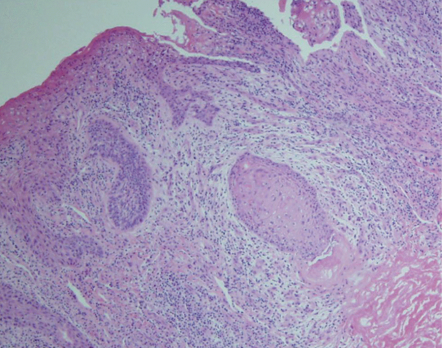
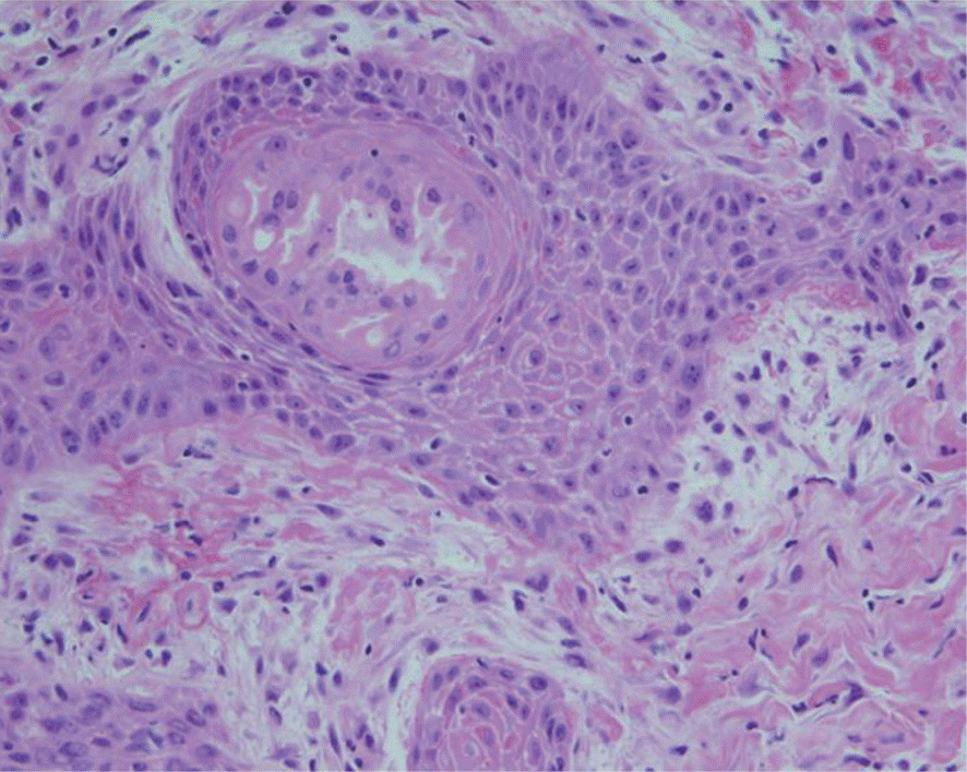
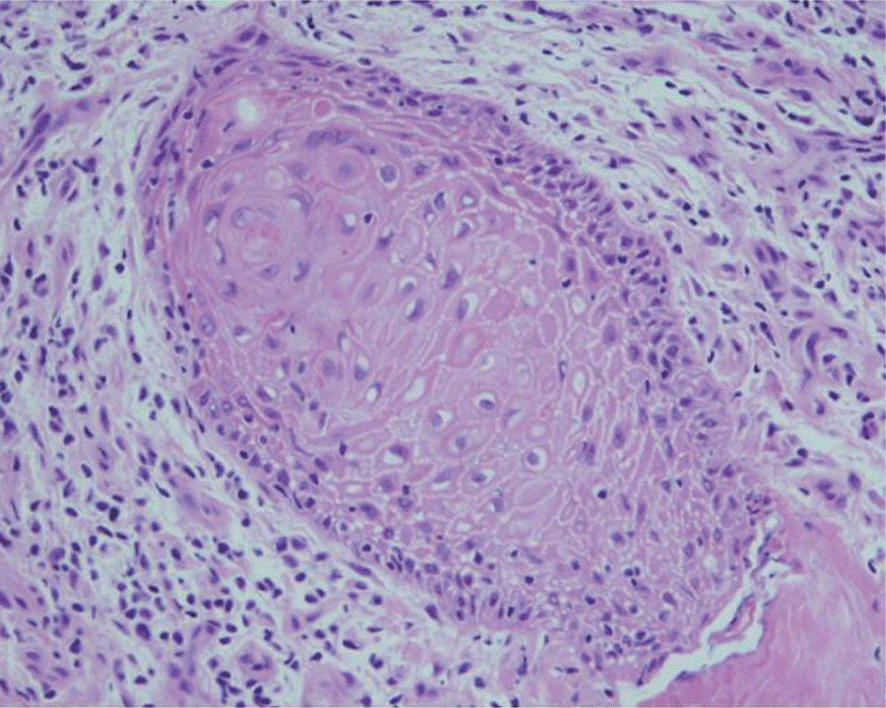
Basosquamous carcinoma is a rare epithelial neoplasm, mostly occurring on the head and neck area. There are few reports of basosquamous carcinoma on the finger. Here, the authors experienced treatment of basosquamous carcinoma on the finger in a radiologist. Treatment was successful by the wide excision and the cross-finger flap operation with a split-thickness skin graft and K-wire fixation. The rare finger basosquamous carcinoma case in our study is likely to be linked with radiation. Considering of the high reliance of C-arm during hand surgeries, we think that the hand of the surgeons should be more strictly protected.
REFERENCES
1. Wermker K, Roknic N, Goessling K, Klein M, Schulze HJ, Hallermann C. Basosquamous carcinoma of the head and neck: clinical and histologic characteristics and their impact on disease progression. Neoplasia. 2015; 17:301–5.

2. Leibovitch I, Huilgol SC, Selva D, Richards S, Paver R. Basosquamous carcinoma: treatment with Mohs micrographic surgery. Cancer. 2005; 104:170–5.
3. Skaria AM. Recurrence of basosquamous carcinoma after Mohs micrographic surgery. Dermatology. 2010; 221:352–5.

4. Giordano BD, Baumhauer JF, Morgan TL, Rechtine GR 2nd. Patient and surgeon radiation exposure: comparison of standard and mini-C-arm fluoroscopy. J Bone Joint Surg Am. 2009; 91:297–304.


 PDF
PDF ePub
ePub Citation
 Print
Print


 XML Download