

 PDF
PDF ePub
ePub Citation
 Print
Print


Abstract
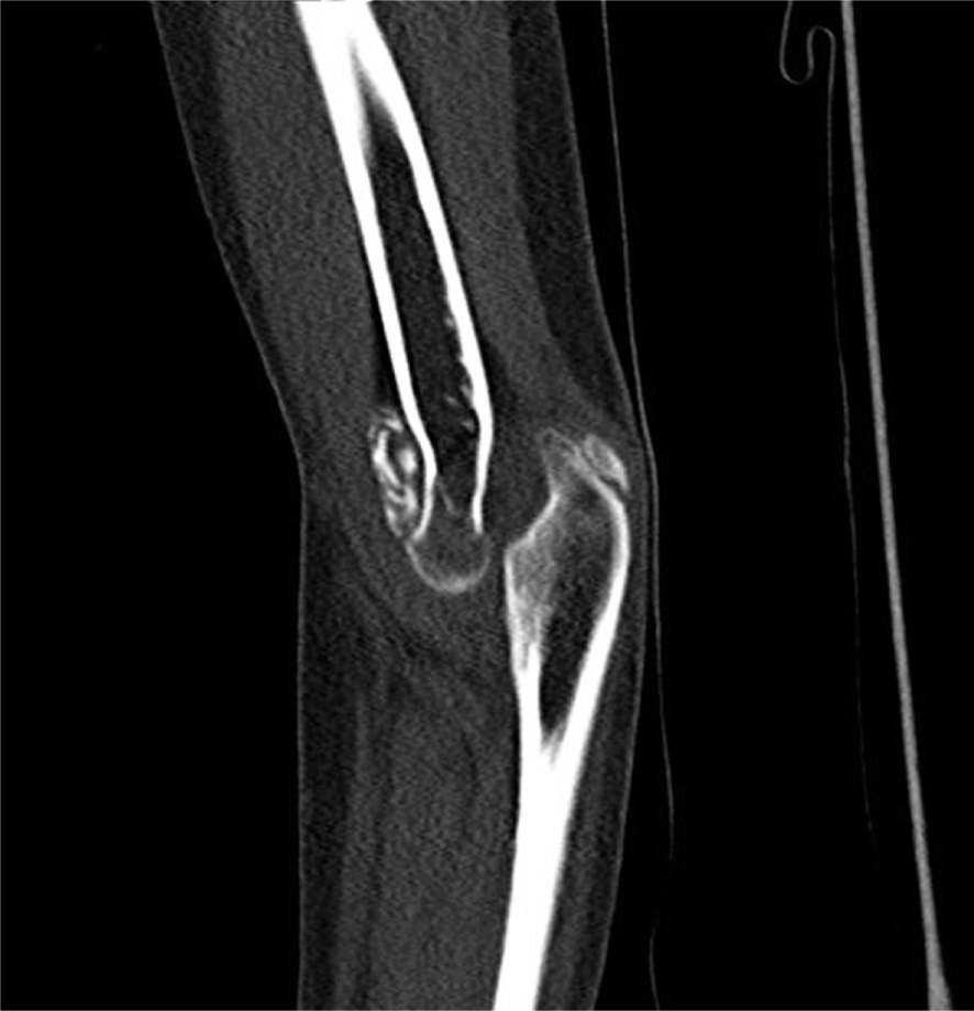
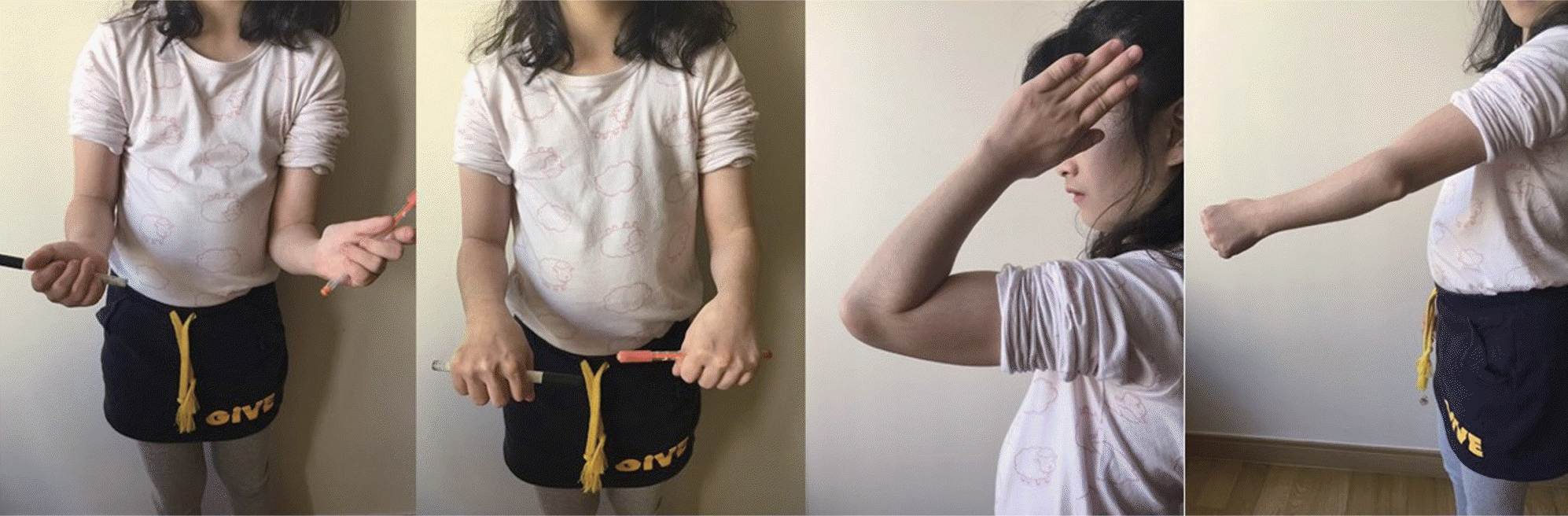
The elbow joint is one of the most inherently stable articulations of the skeleton. Recurrent posterior dislocation of the elbow is a rare condition. We experienced a case of recurrent posterior dislocation of the elbow due to shallow trochlear notch and chronic radial head dislocation that was treated by transplantation of the biceps tendon to the coronoid process. We report on the case with a literature review.
Go to : 
REFERENCES
1. Linscheid RL, Wheeler DK. Elbow dislocations. JAMA. 1965; 194:1171–6.

2. Alcid JG, Ahmad CS, Lee TQ. Elbow anatomy and structural biomechanics. Clin Sports Med. 2004; 23:503–17.
3. Cohen MS, Bruno RJ. The collateral ligaments of the elbow: anatomy and clinical correlation. Clin Orthop Relat Res. 2001; (383):123–30.
4. Shiba R, Sorbie C, Siu DW, Bryant JT, Cooke TD, Wevers HW. Geometry of the humeroulnar joint. J Orthop Res. 1988; 6:897–906.
5. Kapandji IA. The physiology of the joints. Edinburgh: Churchill Livingstone;1982.
6. Cage DJ, Abrams RA, Callahan JJ, Botte MJ. Soft tissue attachments of the ulnar coronoid process. An anatomic study with radiographic correlation. Clin Orthop Relat Res. 1995; (320):154–8.
7. King T. Recurrent dislocation of the elbow. J Bone Joint Surg Br. 1953; 35:50–4.
8. Osborne G, Cotterill P. Recurrent dislocation of the elbow. J Bone Joint Surg Br. 1966; 48:340–6.
9. Milch H. Bilateral recurrent dislocation of the ulna at the elbow. J Bone Joint Surg. 1936; 18:777–80.
10. Wainwright D. Recurrent dislocation of the elbow-joint. Proc R Soc Med. 1947; 40:885–6.
Go to :
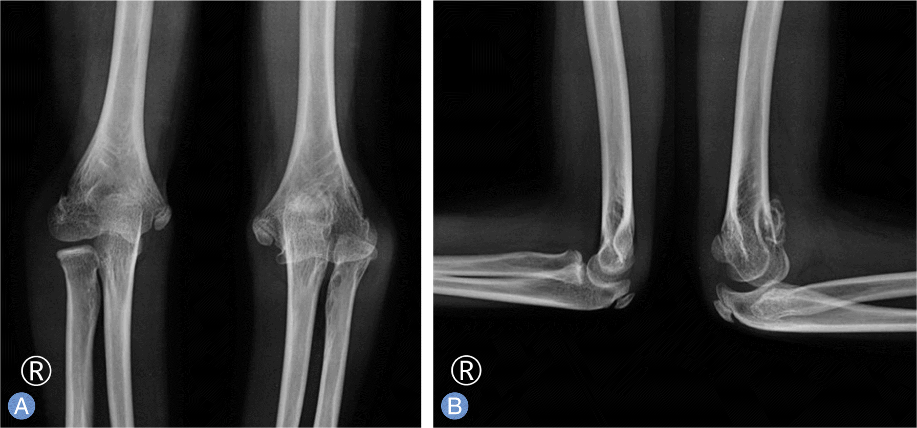 | Fig. 1.Preoperative radiographs. (A) Anteroposterior radiograph showing cubitus varus deformity and chronic radial head dislocation at the left elbow. (B) Lateral radiograph in 90° flexion showing nearly normal articulation of humeroulnar joint and shallow trochlea notch at the left elbow. |

 XML Download