

 PDF
PDF ePub
ePub Citation
 Print
Print


Abstract
Isolated fracture of the distal humeral trochlea occurs while the axial load delivered to the elbow passes through the trochlear of the distal humerus. It has been rarely reported, because of those reasons. The trochlea is located deep inside of the elbow joint space and since it does not have the direct attachment with muscles or ligaments, a force is hardly transmitted directly. Also ulno-humeral joint is less influenced by compressive or shear force than radio-humeral joint. We report a case of isolated trochlear fracture with review of the literature.
REFERENCES
1. Foulk DA, Robertson PA, Timmerman LA. Fracture of the trochlea. J Orthop Trauma. 1995; 9:530–2.
2. Sen RK, Tripahty SK, Goyal T, Aggarwal S. Coronal shear fracture of the humeral trochlea. J Orthop Surg (Hong Kong). 2013; 21:82–6.

3. Abbassi N, Abdeljaouad N, Daoudi A, Yacoubi H. Isolated fracture of the humeral trochlea: a case report and review of the literature. J Med Case Rep. 2015; 9:121.
4. Park JH, Kim JH, Lee JH, Cha SD, Yoo JH. Isolated fracture of the trochlea in humerus: a case report. J Korean Soc Surg Hand. 2006; 11:197–200.
5. Kwan MK, Khoo EH, Chua YP, Mansor A. Isolated displaced fracture of humeral trochlea: a report of two rare cases. Injury Extra. 2007; 38:461–5.
6. Gomati A, Domos P, Crossman P. Delayed surgical management of an isolated trochlear fracture of the elbow. Ann R Coll Surg Engl. 2016; 98:e31–3.
7. Lechasseur B, Laflamme M, Leclerc A, Bedard AM. Incipient malunion of an isolated humeral trochlea fracture treated with an elbow hemiarthroplasty: case report. J Hand Surg Am. 2015; 40:271–5.
8. Worrell RV. Isolated, displaced fracture of the trochlea. N Y State J Med. 1971; 71:2314–5.
9. Goncalves LB, Ring DC. Fractures of the humeral trochlea: case presentations and review. J Shoulder Elbow Surg. 2016; 25:e151–5.
10. Dubberley JH, Faber KJ, Macdermid JC, Patterson SD, King GJ. Outcome after open reduction and internal fixation of capitellar and trochlear fractures. J Bone Joint Surg Am. 2006; 88:46–54.
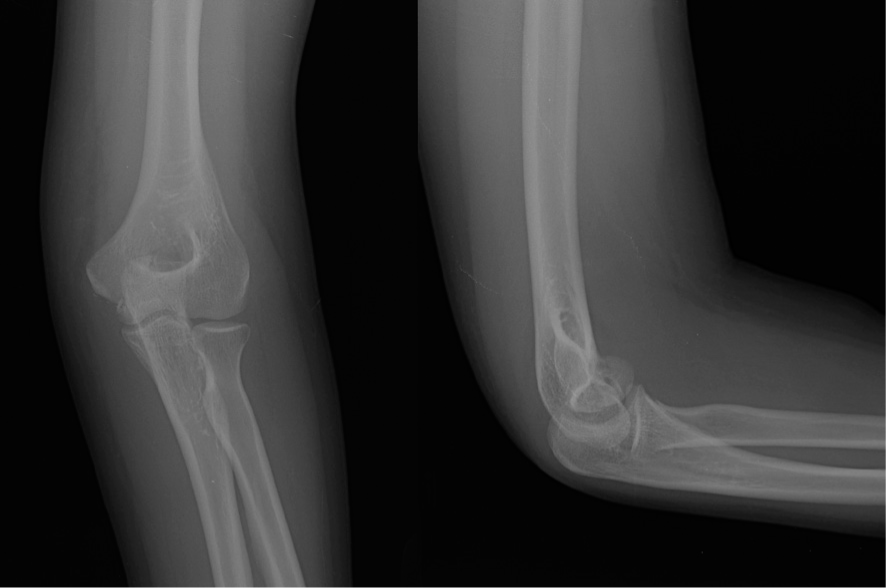
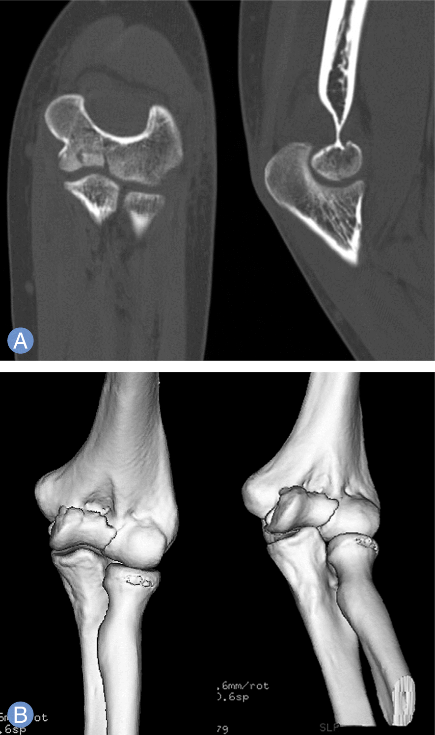
Fig. 3.
(A, B) Postoperative radiograph and sagittal plane views of computed tomography scan shows open reduction and internal fixation with cancellous screw.
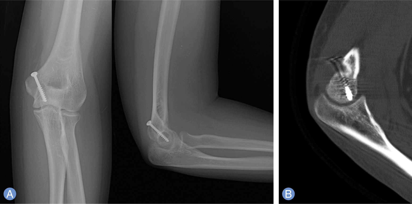
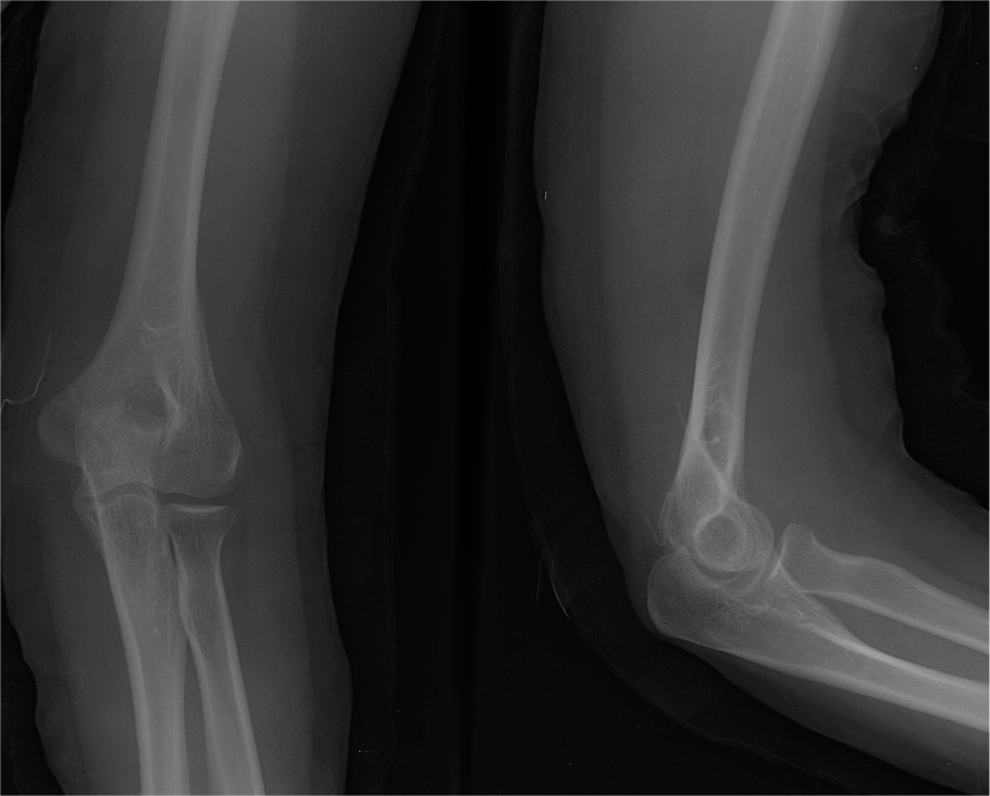
Table 1.
Reports of isolated trochlear fracture
 XML Download