

 PDF
PDF ePub
ePub Citation
 Print
Print


Abstract
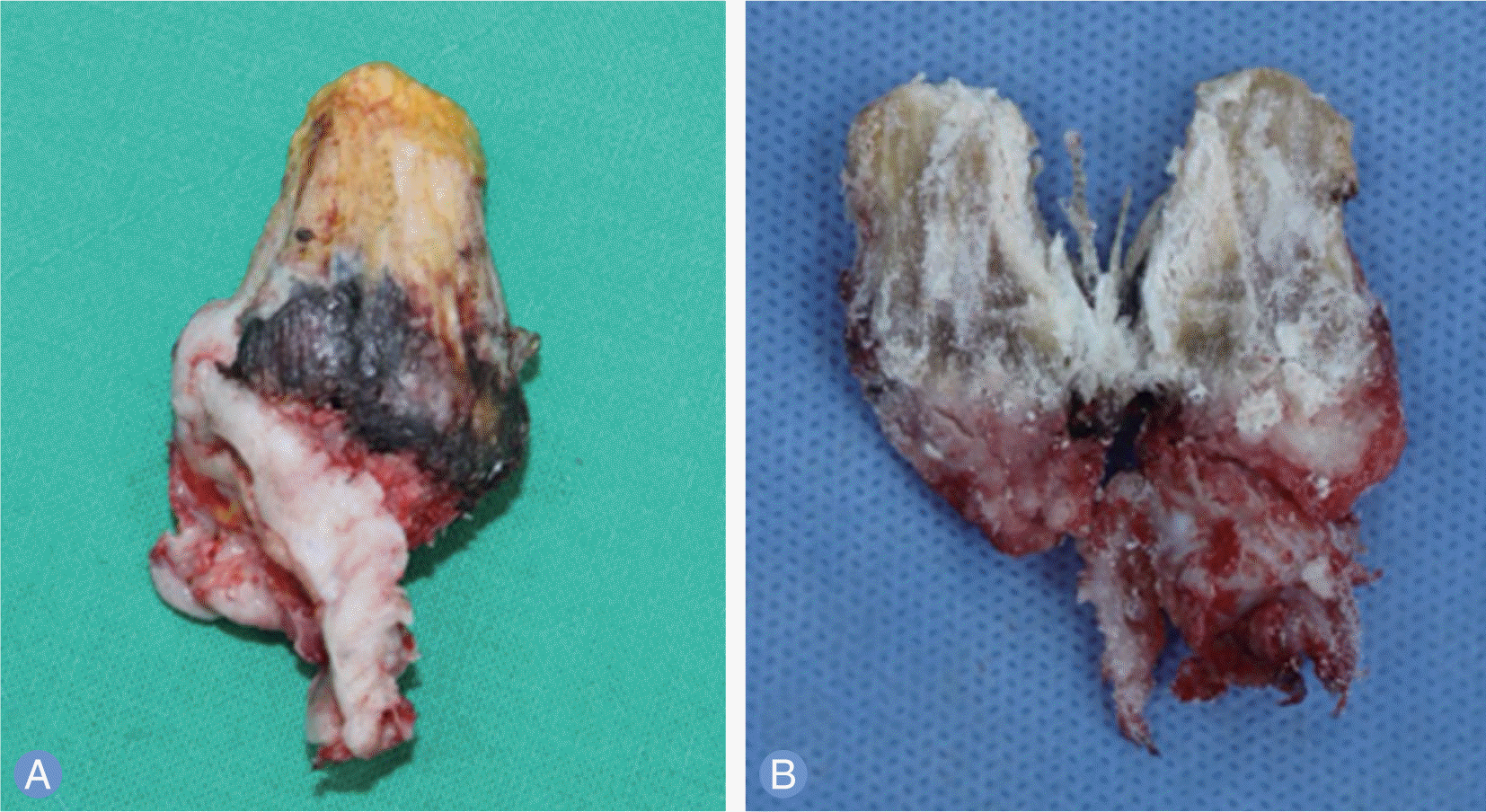
A 81-year-old female presented with a giant cutaneous horn (1.5×2×4 cm sized) over the right hand, rapid growth recent 6 months. Cutaneous horn was excised and split thickness skin graft of the defect was done under regional anesthesia. Histopathology showed overlying a wart. In the current study, we report the case of a patient with giant wart as a giant cutaneous horn in a dorsal side of hand.
Go to : 
REFERENCES
1. Bondeson J. Everard Home, John Hunter, and cutaneous horns: a historical review. Am J Dermatopathol. 2001; 23:362–9.
2. Fernandes NF, Sinha S, Lambert WC, Schwartz RA. Cutaneous horn: a potentially malignant entity. Acta Dermatovenerol Alp Pannonica Adriat. 2009; 18:189–93.
3. Solivan GA, Smith KJ, James WD. Cutaneous horn of the penis: its association with squamous cell carcinoma and HPV-16 infection. J Am Acad Dermatol. 1990; 23:969–72.

4. Cockerell CJ, Larsen F. Benign epithelial tumors and proliferation. Sterry W, Paus R, Burgdorf WHC, editors. Dermatology. 2nd ed.New York: Thieme;2006. p. 1675–6.
5. Yu RC, Pryce DW, Macfarlane AW, Stewart TW. A histopathological study of 643 cutaneous horns. Br J Dermatol. 1991; 124:449–52.
6. Mencia-Gutierrez E, Gutierrez-Diaz E, Redondo-Marcos I, Ricoy JR, Garcia-Torre JP. Cutaneous horns of the eyelid: a clinicopathological study of 48 cases. J Cutan Pathol. 2004; 31:539–43.
7. Copcu E, Sivrioglu N, Culhaci N. Cutaneous horns: are these lesions as innocent as they seem to be? World J Surg Oncol. 2004; 2:18.
8. Kumar S, Bijalwan P, Saini SK. Carcinoma buccal mucosa underlying a giant cutaneous horn: a case report and review of the literature. Case Rep Oncol Med. 2014; 2014:518372.
9. Mantese SA, Diogo PM, Rocha A, Berbert AL, Ferreira AK, Ferreira TC. Cutaneous horn: a retrospective histopathological study of 222 cases. An Bras Dermatol. 2010; 85:157–63.
10. Lowe FC, McCullough AR. Cutaneous horns of the penis: an approach to management: case report and review of the literature. J Am Acad Dermatol. 1985; 13:369–73.
Go to :
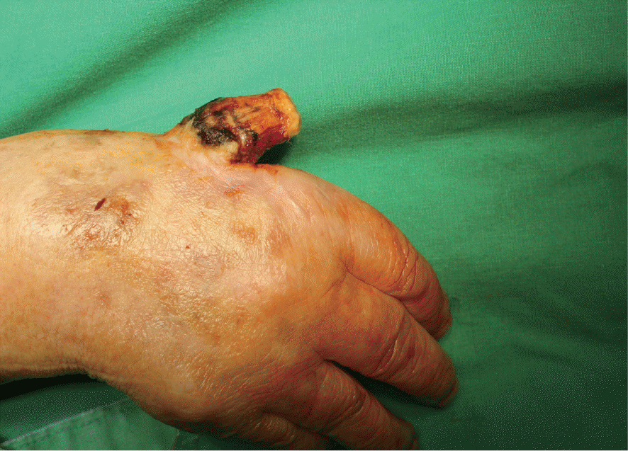 | Fig. 1.A horny growth of around 4 cm in length with a broad base over the dorsal aspect of the right hand. |
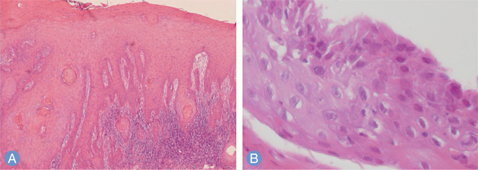 | Fig. 3.The histologic characteristics of this lesion is epidermal hyperplasia manifested by hyperkeratosis, parakeratosis, acanthosis, papillomatosis and elongation of rete ridges (H&E, x40) (A), Koilocytotic cells, characterized by small, round, deeply basophilic nuclei surrounded by a clear halo and pale-staining cytoplasm are also present (H&E, x400) (B).
|
 XML Download