Abstract
Compartment syndrome is caused by elevated pressure in a restricted compartment. It typically occurs after fractures of the extremities and usually has an acute clinical progression. Chronic compartment syndrome is another relatively well known form, typically associated with forceful exercise. Also, there are various reports of compartment syndrome not associated with typical causes. However, reports on compartment syndrome with unknown etiology are rare and there has been none in Korean literature. We report a case of compartment syndrome with no recognizable cause, hence classified as idiopathic.
Go to : 
References
1. Duckworth AD, Mitchell SE, Molyneux SG, White TO, Court-Brown CM, McQueen MM. Acute compartment syndrome of the forearm. J Bone Joint Surg Am. 2012; 94:e63.

2. Brown JS, Wheeler PC, Boyd KT, Barnes MR, Allen MJ. Chronic exertional compartment syndrome of the forearm: a case series of 12 patients treated with fasciotomy. J Hand Surg Eur Vol. 2011; 36:413–9.

3. Matziolis G, Erli HJ, Rau MH, Klever P, Paar O. Idiopathic compartment syndrome: a case report. J Trauma. 2002; 53:122–4.

4. Smith K, Wolford RW. Acute idiopathic compartment syndrome of the forearm in an adolescent. West J Emerg Med. 2015; 16:158–60.

5. Jose RM, Viswanathan N, Aldlyami E, Wilson Y, Moiemen N, Thomas R. A spontaneous compartment syndrome in a patient with diabetes. J Bone Joint Surg Br. 2004; 86:1068–70.

6. Boody AR, Wongworawat MD. Accuracy in the measurement of compartment pressures: a comparison of three commonly used devices. J Bone Joint Surg Am. 2005; 87:2415–22.

7. Whitesides TE, Heckman MM. Acute compartment syndrome: update on diagnosis and treatment. J Am Acad Orthop Surg. 1996; 4:209–18.

Go to : 
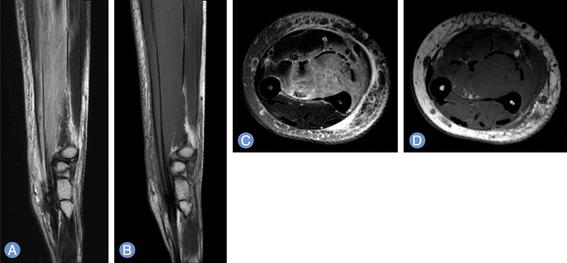
 | Fig. 1.Sagittal T2-weighted fat supression (A), sagittal T1-weighted (B), axial T2-weighted fat supression (C), axial T1-weighted (D) magnetic resonance image of the patient. The fat suppressed T2-weighted magnetic resonance image demonstrating increased signal of flexor muscle groups especially involving deep layer of entire forearm with dorsal bulging of interosseous membrane between ulna and radius and T1-weighted image shows similar signal intensity to that of normal muscle. |
Table 1.
Etiology of compartment syndrome


 PDF
PDF ePub
ePub Citation
 Print
Print


 XML Download