Abstract
Purpose:
The goal of this retrospective study is to compare radiologic outcome and clinical outcome between operative and non-operative treatment of unstable distal radius fracture in patients over 65-year-old.
Methods:
From December 2006 to December 2011, 114 patients over 65-year-old were enrolled in the present study. 45 patients underwent non-operative treatment, and 69 patients underwent operative treatment. We retrospectively reviewed radiologic results and clinical results and then compared the two groups. Radiologic results include radial inclination (RI), volar tilt angle (VT) and radial shortening (RS) shown on the last radiograph and clinical results including disabilities of the arm, shoulder and hand (DASH) scores, modified Mayo wrist score (MMWS), and range of motion (ROM) of wrist.
Results:
All cases presented bone-union. Among the patients who received non-operative treatments, average RI of 15.5°, average VT of 14.1°, average RS of 5.3 mm, The patients who received operative treatments showed average volar tilt of 3.9°, average VT of 18.2°, and average RS of 1.1 mm. RS showed a significant difference (p<0.05). At Clinical evaluation, DASH score, MMWS score, the ROM of wrist joint did not show significant difference (p>0.05).
Go to : 
References
1. Abbaszadegan H, Jonsson U. External fixation or plaster cast for severely displaced Colles' fractures? Prospective 1-year study of 46 patients. Acta Orthop Scand. 1990; 61:528–30.

2. Abbaszadegan H, Jonsson U. von Sivers K. Prediction of instability of Colles' fractures. Acta Orthop Scand. 1989; 60:646–50.
3. Alffram PA, Bauer GC. Epidemiology of fractures of the forearm: a biomechanical investigation of bone strength. J Bone Joint Surg Am. 1962; 44:105–14.
4. Anzarut A, Johnson JA, Rowe BH, Lambert RG, Blitz S, Majumdar SR. Radiologic and patient-reported functional outcomes in an elderly cohort with conservatively treated distal radius fractures. J Hand Surg Am. 2004; 29:1121–7.

5. Azzopardi T, Ehrendorfer S, Coulton T, Abela M. Unstable extra-articular fractures of the distal radius: a prospective, randomised study of immobilisation in a cast versus supplementary percutaneous pinning. J Bone Joint Surg Br. 2005; 87:837–40.
6. Barton T, Chambers C, Bannister G. A comparison between subjective outcome score and moderate radial shortening following a fractured distal radius in patients of mean age 69 years. J Hand Surg Eur Vol. 2007; 32:165–9.

7. Beumer A, McQueen MM. Fractures of the distal radius in low-demand elderly patients: closed reduction of no value in 53 of 60 wrists. Acta Orthop Scand. 2003; 74:98–100.

8. Boyd LG, Horne JG. The outcome of fractures of the distal radius in young adults. Injury. 1988; 19:97–100.

9. Christensen OM, Christiansen TC, Krasheninnikoff M, et al. Plaster cast compared with bridging external fixation for distal radius fractures of the Colles' type. Int Orthop. 2001; 24:358–60.

10. Jaremko JL, Lambert RG, Rowe BH, Johnson JA, Majumdar SR. Do radiographic indices of distal radius fracture reduction predict outcomes in older adults receiving conservative treatment? Clin Radiol. 2007; 62:65–72.
11. Ark J, Jupiter JB. The rationale for precise management of distal radius fractures. Orthop Clin North Am. 1993; 24:205–10.

12. Howard PW, Stewart HD, Hind RE, Burke FD. External fixation or plaster for severely displaced comminuted Colles’ fractures? A prospective study of anatomical and functional results. J Bone Joint Surg Br. 1989; 71:68–73.

13. McQueen M, Caspers J. Colles fracture: does the anatomical result affect the final function? J Bone Joint Surg Br. 1988; 70:649–51.

14. Makhni EC, Ewald TJ, Kelly S, Day CS. Effect of patient age on the radiographic outcomes of distal radius fractures subject to nonoperative treatment. J Hand Surg Am. 2008; 33:1301–8.

15. Young BT, Rayan GM. Outcome following nonoperative treatment of displaced distal radius fractures in low-demand patients older than 60 years. J Hand Surg Am. 2000; 25:19–28.

16. Arora R, Lutz M, Deml C, Krappinger D, Haug L, Gabl M. A prospective randomized trial comparing nonoperative treatment with volar locking plate fixation for displaced and unstable distal radial fractures in patients sixty-five years of age and older. J Bone Joint Surg Am. 2011; 93:2146–53.

17. Egol KA, Walsh M, Romo-Cardoso S, Dorsky S, Paksima N. Distal radial fractures in the elderly: operative compared with nonoperative treatment. J Bone Joint Surg Am. 2010; 92:1851–7.

18. Lidstrom A. Fractures of the distal end of the radius: a clinical and statistical study of end results. Acta Orthop Scand Suppl. 1959; 41:1–118.
19. Frykman G. Fracture of the distal radius including sequelae: shoulder-hand-finger syndrome, disturbance in the distal radio-ulnar joint and impairment of nerve function: a clinical and experimental study. Acta Orthop Scand. 1967; (Suppl 108):3+.
20. Knirk JL, Jupiter JB. Intra-articular fractures of the distal end of the radius in young adults. J Bone Joint Surg Am. 1986; 68:647–59.

21. Rodriguez-Merchan EC. Management of comminuted fractures of the distal radius in the adult: conservative or surgical? Clin Orthop Relat Res. 1998; (353):53–62.
Go to : 
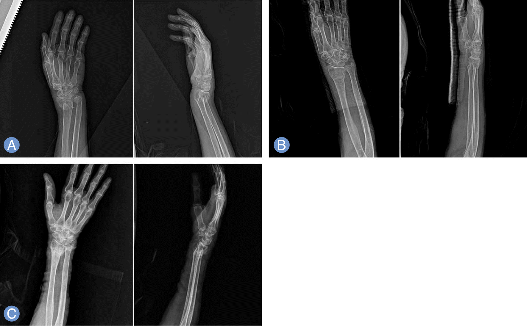
 | Fig. 1.Radiographs of 84-yerar-old female who received conservative treatment. (A) Radiograph shows 1.8mm radial shortening and 24.7° dorsal tilting. (B) Closed reduction and sugar tong splint was applied. Radial shortening reduced to 0 mm and dorsal tilting decreased to 6.4°. (C) Twelve month follow-up radiographs shows 1.2 mm radial shortening and 18.5° dorsal tilting. |
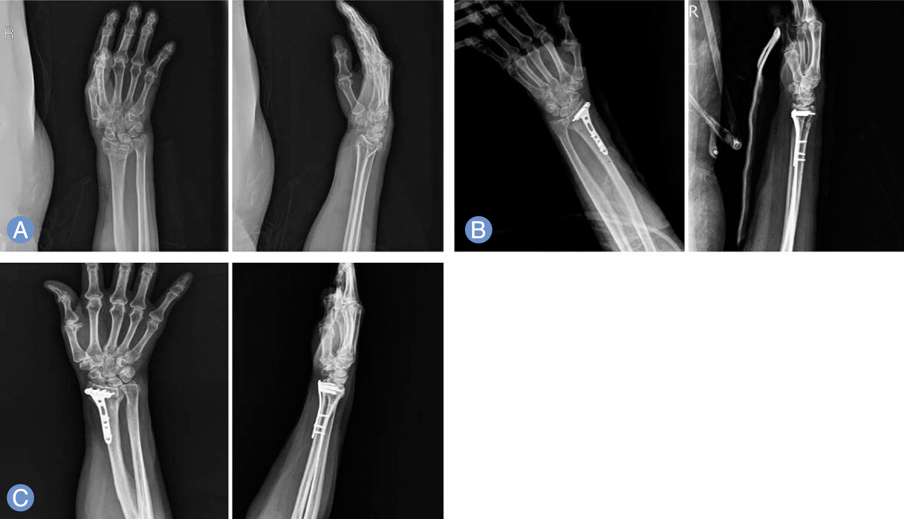
 | Fig. 2.Radiographs of 76-yerar-old female who underwent operative treatment. (A) Preoperative radiograph shows 5.4 mm radial shortening and 11.7° dorsal tilting. (B) Open reduction and volar locking plate was applied. Radial shortening reduced to 0mm and dorsal tilting decreased to 2.2°. (C) Seven month follow-up radiographs shows 0.5 mm radial shortening and 4.8° dorsal tilting. |
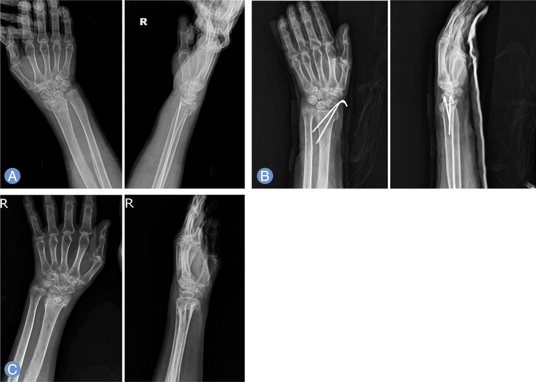
 | Fig. 3.Radiographs of 72-yerar-old female who underwent operative treatment. (A) Preoperative radiograph shows 2.0 mm radial shortening and 4.8° dorsal tilting. (B) Closed reduction and K-wire fixation was done. Radial shortening reduced to 1.7 mm and dorsal tilting decreased to 4.5°. (C) Eight month follow-up radiographs shows 2 mm radial shortening and 3.1° dorsal tilting. |
Table 1.
Summary of dermographic data
Table 2.
Complications of distal radius fracture for both
Table 3.
Radiologic measurements for both groups
Table 4.
Functional outcomes at final follow-up for both groups


 PDF
PDF ePub
ePub Citation
 Print
Print


 XML Download