Abstract
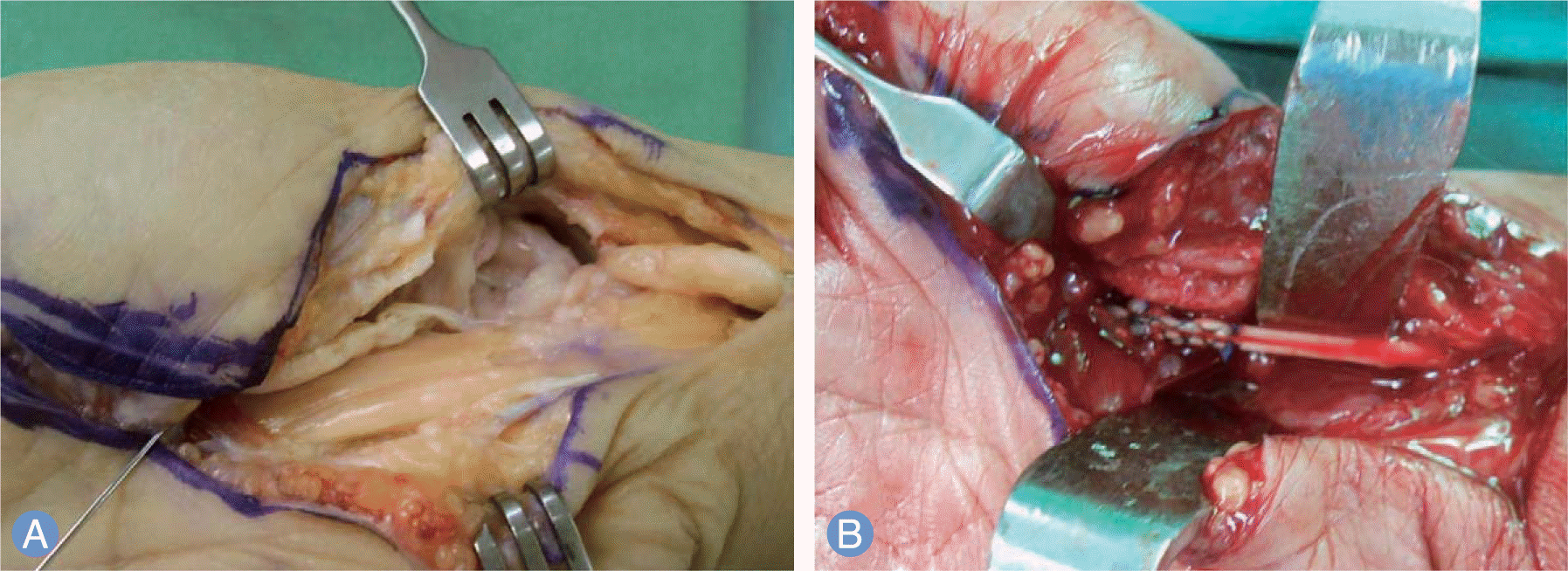
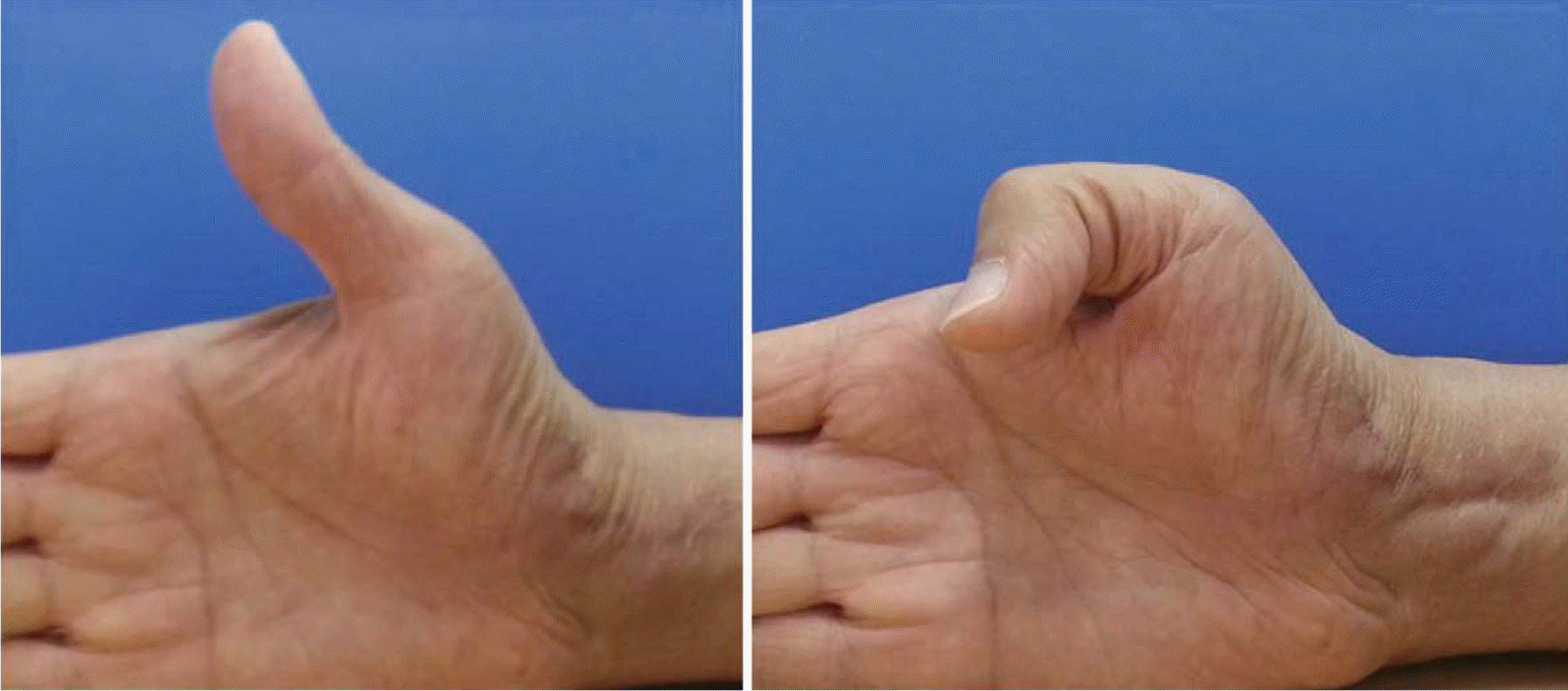
Flexor pollicis longus rupture due to scaphoid nonunion is very rare complication. It has never been reported in the Korean literatures. We reported a case of flexor pollicis longus rupture due to scaphoid non union that was treated by tendon graft with palmaris longus and osteosynthesis with bone graft.
Go to : 
References
1. Mahring M, Semple C, Gray IC. Attritional flexor tendon rupture due to a scaphoid non union imitating an anterior interosseous nerve syndrome: a case report. J Hand Surg Br. 1985; 10:62–4.
2. Cross AB. Rupture of the flexor pollicis longus tendon resulting from the non-union of a scaphoid fracture. J Hand Surg Br. 1988; 13:80–2.

3. Thomsen S, Falstie-Jensen S. Rupture of the flexor pollicis longus tendon associated with an ununited fracture of the scaphoid. J Hand Surg Am. 1988; 13:220–2.

4. McLain RF, Steyers CM. Tendon ruptures with scaphoid nonunion: a case report. Clin Orthop Relat Res. 1990; (255):117–20.
5. Zachee B, De Smet L, Fabry G. Flexor pollicis longus rupture with scaphoid nonunion: a case report and literature study. Acta Orthop Belg. 1991; 57:456–8.
6. Saitoh S, Hata Y, Murakami N, Nakatsuchi Y, Seki H, Takaoka K. Scaphoid nonunion and flexor pollicis longus tendon rupture. J Hand Surg Am. 1999; 24:1211–9.

7. Yamazaki H, Kato H, Hata Y, Nakatsuchi Y, Tsuchikane A. Closed rupture of the flexor tendons caused by carpal bone and joint disorders. J Hand Surg Eur Vol. 2007; 32:649–53.

8. Wacker J, McKie S, MacLean JG. Delayed sequential ruptures of the index and thumb flexor tendons due to an occult scaphoid nonunion. J Hand Surg Br. 1999; 24:741–3.

9. Kakarala G, Arya AP, Compson JP. Extensor pollicis longus tendon rupture due to scaphoid non-u. J Hand Surg Br. 2006; 31:353.
10. Mannerfelt L, Norman O. Attrition ruptures of flexor tendons in rheumatoid arthritis caused by bony spurs in the carpal tunnel: a clinical and radiological study. J Bone Joint Surg Br. 1969; 51:270–7.
Go to : 
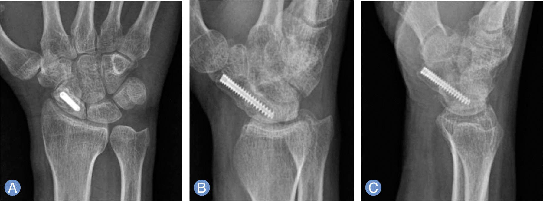
 | Fig. 1.
(A-C) Preoperative radiography. Scaphoid nonunion with humpback deformity is observed. The distal fragment is located anterior to the proximal one. |


 PDF
PDF ePub
ePub Citation
 Print
Print


 XML Download