Abstract
Indicators for local steroid injection on the hands include trigger finger, De Quervain’s disease, carpal tunnel syndrome and trapeziometacarpal joint arthritis. Local steroid injection is an effective technique for rapid alleviation of symptoms and return to daily life. Complications following local steroid injection include depigmentation of the skin, subcutaneous fat atrophy, infection and tendon rupture. Tendon rupture and infection rarely occur as severe complications, and local steroid injection should not be abused or misused. The authors experienced a rupture of the extensor mechanism at extensor zone V after repeated local steroid injection to treat vague pain in the second metacarpophalangeal joint, followed by reconstruction of the extensor mechanism through tendon transfer and sagittal band reconstruction. We herein report a case with the literature review.
Go to : 
References
1. Dean BJ, Lostis E, Oakley T, Rombach I, Morrey ME, Carr AJ. The risks and benefits of glucocorticoid treatment for tendinopathy: a systematic review of the effects of local glucocorticoid on tendon. Semin Arthritis Rheum. 2014; 43:570–6.

2. Gyuricza C, Umoh E, Wolfe SW. Multiple pulley rupture following corticosteroid injection for trigger digit: case report. J Hand Surg Am. 2009; 34:1444–8.

3. Coombes BK, Bisset L, Vicenzino B. Efficacy and safety of corticosteroid injections and other injections for management of tendinopathy: a systematic review of randomised controlled trials. Lancet. 2010; 376:1751–67.

4. Hugate R, Pennypacker J, Saunders M, Juliano P. The effects of intratendinous and retrocalcaneal intrabursal injections of corticosteroid on the biomechanical properties of rabbit Achilles tendons. J Bone Joint Surg Am. 2004; 86:794–801.

5. Balasubramaniam P, Prathap K. The effect of injection of hydrocortisone into rabbit calcaneal tendons. J Bone Joint Surg Br. 1972; 54:729–34.

Go to : 
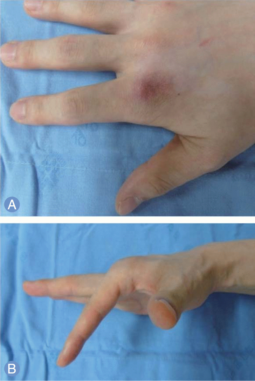
 | Fig. 1.Photographs of hand before surgery. (A) Erythematous skin color change on dorsal aspect of second metacarpophalangeal joint. (B) Loss of active extension at the second metacarpophalangeal joint. |
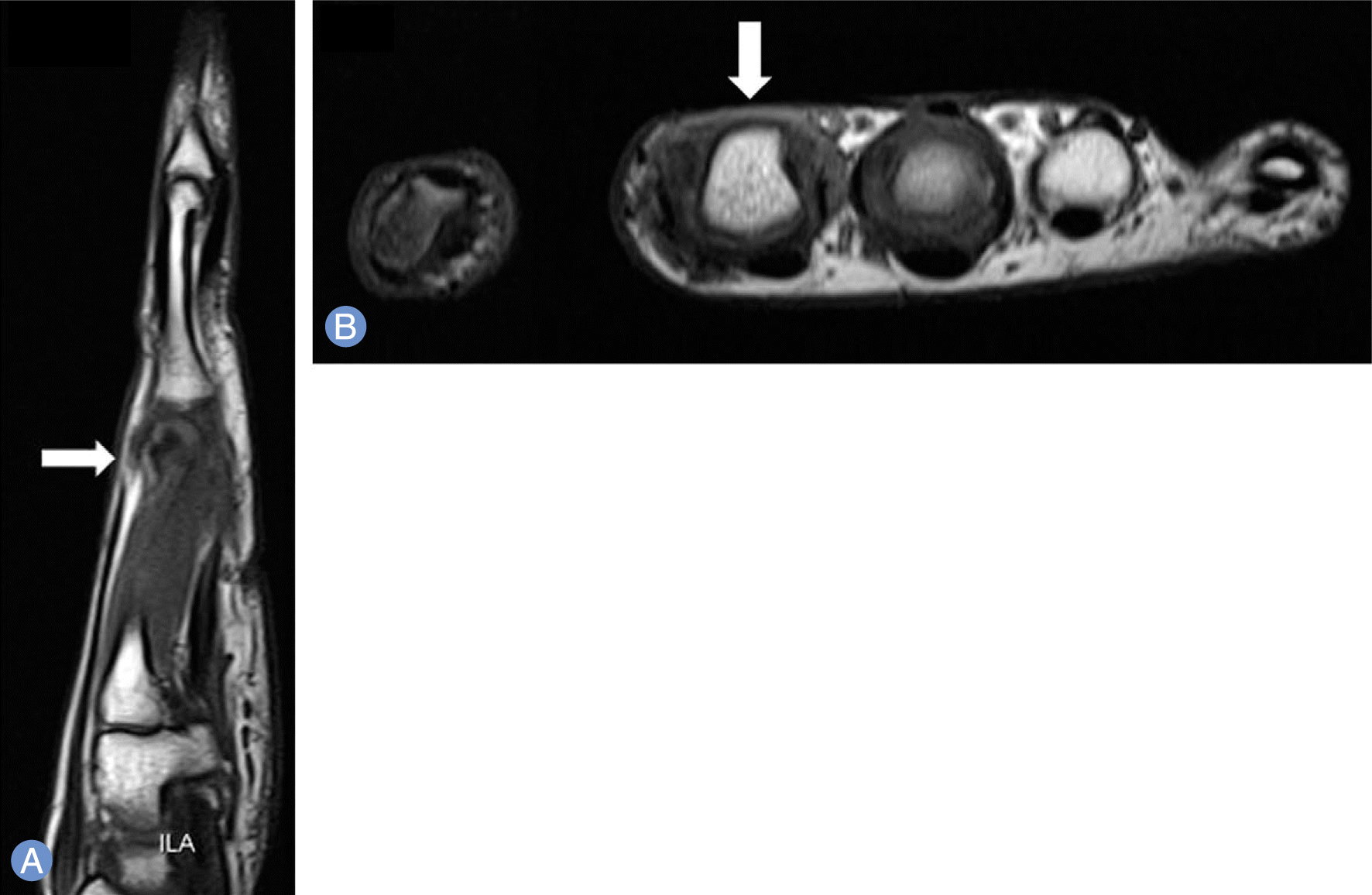
 | Fig. 2.Magnetic resonance images. (A) T1-weight sagittal image demonstrates loss of continuity of extensor tendon rupture at the second metacarpophalangeal joint and proximal stump retraction by approximately 2 cm (arrow). (B) T1-weight axial image demonstrates a defect of the extensor tendon and sagittal band around the second metacarpophalangeal joint (arrow). |
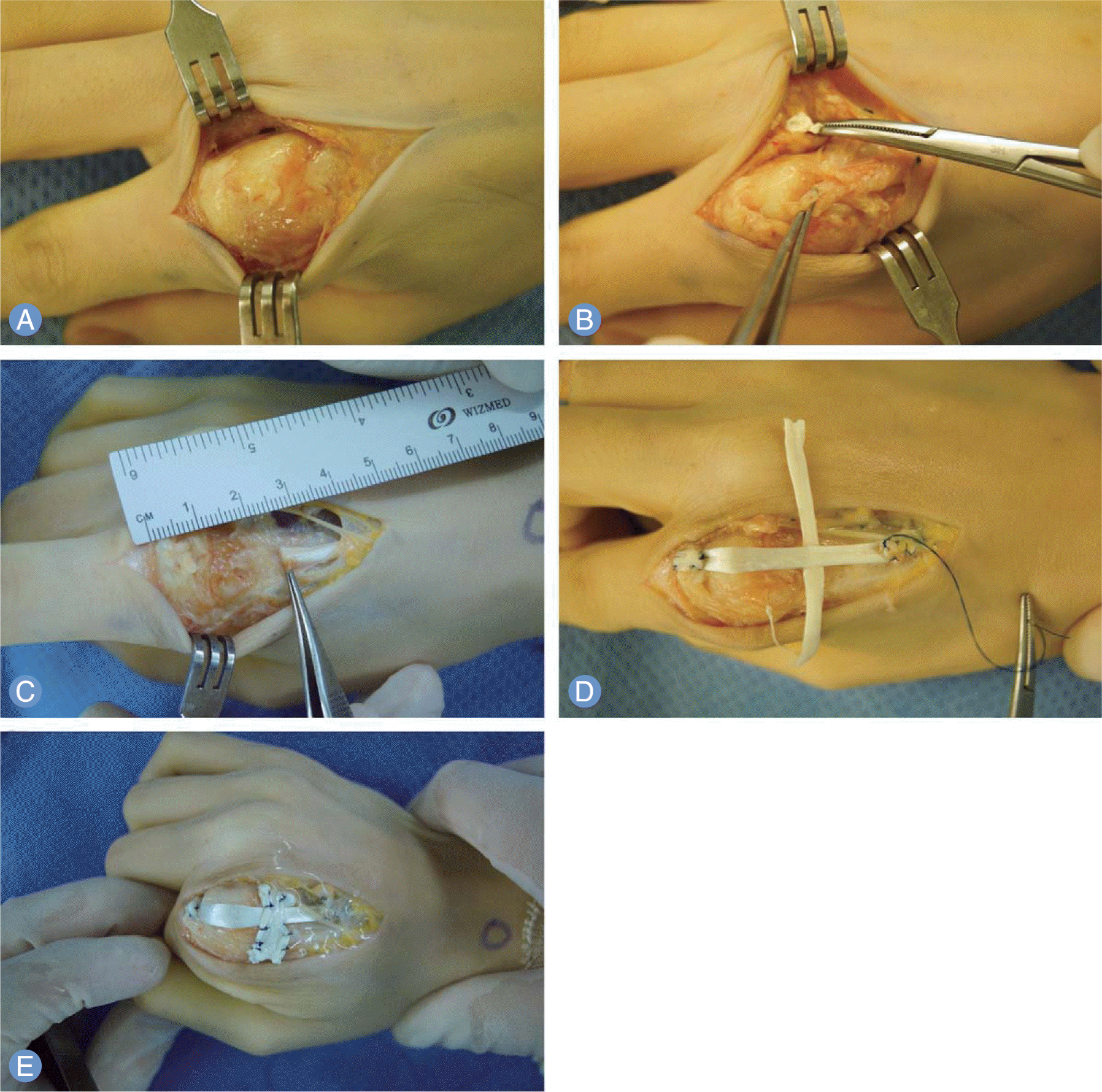
 | Fig. 3.Photographs of operation. (A) Defect of extensor mechanism at zone V with scar tissues and granulation tissues. There are loss of continuity of the extensor digitorum communis tendons and extensor indicis proprius tendon of the index finger at zone V. Additionally, defect of the sagittal band is demonstrated. (B) Tendon ends have frayed, necrotic, adhesive and degenerative appearance with foreign body deposition which is suggested to be steroid. (C) Tendon gap is approximately 2.5 cm. (D) Reconstruction of the extensor tendon with autologous palmaris longus tendon graft. (E) Reconstruction of sagittal band using palmaris longus tendon to prevent subluxation of reconstructed extensor tendon. |


 PDF
PDF ePub
ePub Citation
 Print
Print


 XML Download