

 PDF
PDF ePub
ePub Citation
 Print
Print


Abstract
Purpose:
Our purpose was to assess the results of lunate excision and tendon ball implantation with temporary scaphocapitate fixation for Lichtman stage IIIB Kienböck’s disease in middle-aged patients.
Methods:
Ten patients with Lichtman stage IIIB Kienböck’s disease who underwent lunate excision and tendon ball implantation and followed up at least 24 months were analyzed. There were 4 males and 6 females. The mean age at the time of surgery was 55.4 years (range, 48-67 years), and follow-up period ranged from 24 to 68 months (mean, 46 months). Radiocarpal joint pain, grip strength, return to daily living activity, range of motion were evaluated and radiologic findings of preoperative, postoperative and last follow-up were evaluated.
Results:
All patients returned to daily living activity after 6 months of surgery. At the last follow up, 8 patients had no pain and 2 patients experienced mild pain occasionally. The mean improvement of extension arc was 14.5°, and the mean flexion arc improved 8.5°. The mean grip strength was 88% of unaffected side. The mean carpal height ratio was 0.49 preoperatively, 0.47 at final followup. No patients showed osteoarthritis change at the last follow-up. The mean Cooney’s wrist function were 83, 4 patients had excellent, 4 had good, and 2 had fair.
Go to : 
References
1. Lutsky K, Beredjiklian PK. Kienbock disease. J Hand Surg Am. 2012; 37:1942–52.
2. Jensen CH. Intraosseous pressure in Kienbock’s disease. J Hand Surg Am. 1993; 18:355–9.
3. Gelberman RH, Salamon PB, Jurist JM, Posch JL. Ulnar variance in Kienbock’s disease. J Bone Joint Surg Am. 1975; 57:674–6.

4. Lichtman DM, Degnan GG. Staging and its use in the determination of treatment modalities for Kienbock’s disease. Hand Clin. 1993; 9:409–16.
5. Armistead RB, Linscheid RL, Dobyns JH, Beckenbaugh RD. Ulnar lengthening in the treatment of Kienbock’s disease. J Bone Joint Surg Am. 1982; 64:170–8.
6. Weiss AP, Weiland AJ, Moore JR, Wilgis EF. Radial shortening for Kienbock disease. J Bone Joint Surg Am. 1991; 73:384–91.
7. Moran SL, Cooney WP, Berger RA, Bishop AT, Shin AY. The use of the 4 + 5 extensor compartmental vascularized bone graft for the treatment of Kienbock’s disease. J Hand Surg Am. 2005; 30:50–8.
8. Cheon SJ, Lim JM, Kim HT, Suh JT. Treatment of Kienbock’s disease using the 4+5 extensor compartmental vascularized bone grafting procedure: early experience. J Korean Orthop Assoc. 2010; 45:256–63.
9. Yajima H, Ono H, Tamai S. Temporary internal fixation of the scaphotrapezio-trapezoidal joint for the treatment of Kienbock’s disease: a preliminary study. J Hand Surg Am. 1998; 23:402–10.
10. Yajima H, Tamai S, Mizumoto S, Ono H, Inada Y. Treatment of Kienbock’s disease with vascular bundle implantation and triscaphe arthrodesis. Nakamura R, Linscheid RL, Miura T, editors. Wrist disorders: current concepts and challenges. Tokyo: Springer-Verlag;. 1992; 101–9.
11. Ueba Y, Nosaka K, Seto Y, Ikeda N, Nakamura T. An operative procedure for advanced Kienbock’s disease. Excision of the lunate and subsequent replacement with a tendon-ball implant. J Orthop Sci. 1999; 4:207–15.
12. Kato H, Usui M, Minami A. Long-term results of Kienbock’s disease treated by excisional arthroplasty with a silicone implant or coiled palmaris longus tendon. J Hand Surg Am. 1986; 11:645–53.
13. Kucuk L, Ozdemir O, Coskunol E, Sugun TS, Ozaksar K. The effect of excisional arthroplasty with palmaris longus tendon on carpal height ratio in Stage 3 Kienbock’s disease. Acta Orthop Traumatol Turc. 2011; 45:393–8.
14. Matsuhashi T, Iwasaki N, Kato H, Minami M, Minami A. Clinical outcomes of excision arthroplasty for Kienbock’s disease. Hand Surg. 2011; 16:277–82.
15. Sakai A, Toba N, Oshige T, Menuki K, Hirasawa H, Nakamura T. Kienbock disease treated by excisional arthroplasty with a palmaris longus tendon ball: a comparative study of cases with or without bone core. Hand Surg. 2004; 9:145–9.
16. Zeplin PH, Ziegler UE. Long-term results after resection arthroplasty in Kienbock’s disease. J Hand Surg Eur Vol. 2013; 38:553–4.
17. Takase K, Imakiire A. Lunate excision, capitate osteotomy, and intercarpal arthrodesis for advanced Kienbock disease: long-term follow-up. J Bone Joint Surg Am. 2001; 83:177–83.
18. Peste JL. Discussion. Bull Soc Anat Paris. 1843; 18:169–17.
19. Lee ML. The intraosseus arterial pattern of the carpal lunate bone and its relation to avascular necrosis. Acta Orthop Scand. 1963; 33:43–55.
20. Gelberman RH, Szabo RM. Kienbock’s disease. Orthop Clin North Am. 1984; 15:355–67.
21. Kienböck R. Uber traumatische malazie des mondeins und ihre folgezustande: entartungsformen und korm-pressionsfrakturen. Fortschr gebeite rontgenstr Nuklearmed Erganzungsband O. 1910; 16:79–103.
22. Beckenbaugh RD, Shives TC, Dobyns JH, Linscheid RL. Kienbock’s disease: the natural history of Kienbock’s disease and consideration of lunate fractures. Clin Orthop Relat Res. 1980; (149):98–106.
23. Hulten O. Uber anatomische variationen der handge-lenkknochen. Acta Radiol. 1928; 9:155–68.
24. Gelberman RH, Bauman TD, Menon J, Akeson WH. The vascularity of the lunate bone and Kienbock’s disease. J Hand Surg Am. 1980; 5:272–8.
25. Stahl F. On lunatomalacia (Kienbock’s disease): a clinical and roentgenological study, especially on its pathogenesis and the late results of immobilization treatment. Acta Chir Scand. 1947; 126(Suppl):1–133.
26. Innes L, Strauch RJ. Systematic review of the treatment of Kienbock’s disease in its early and late stages. J Hand Surg Am. 2010; 35:713–7.e14.
27. Swanson AB. Silicone rubber implants for the replacement of the carpal scaphoid and lunate bones. Orthop Clin North Am. 1970; 1:299–309.
28. Carroll RE. Long-term review of fascial replacement after excision of the carpal lunate bone. Clin Orthop Relat Res. 1997; (342):59–63.
29. Horita K, Ikuta Y, Murakami T, Ochi M, Mochizuki Y. An experimental study on the bone-core tendon ball replacement for the treatment of Kienböck’s disease. J Japanese Soc Surg Hand. 1990; 7:767–71.
30. Ishiguro T. Experimental and clinical studies of Kienbock’s disease: excision of the lunate followed by packing of the free tendon. Nihon Seikeigeka Gakkai Zasshi. 1984; 58:509–22.
31. Yajima H, Kobata Y, Yamauchi T, Takakura Y. Advanced Kienbock’s disease treated with implantation of a tendon roll and temporary partial fixation of the wrist. Scand J Plast Reconstr Surg Hand Surg. 2004; 38:340–6.
32. Lee KS, Oh KJ, Yeo WJ, Song HS, Park SJ. Long-term results of limited intercarpal arthrodesis combined with lunate excision and rolled-tendon arthroplasty in advanced Kienbock disease. J Korean Orthop Assoc. 2002; 7:42–7.
33. Yang JY, Shin HD, Ahn SR, Rhee KJ, Lee JK, Choi JH. Scaphocapitate fusion for Kienbock disease. J Korean Soc Fract. 1999; 12:446–51.
34. Mariconda M, Soscia E, Sirignano C, Smeraglia F, Soldati A, Balato G. Long-term clinical results and MRI changes after tendon ball arthroplasty for advanced Kienbock’s disease. J Hand Surg Eur Vol. 2013; 38:508–14.
35. Minami A, Kimura T, Suzuki K. Long-term results of Kienbock’s disease treated by triscaphe arthrodesis and excisional arthroplasty with a coiled palmaris longus tendon. J Hand Surg Am. 1994; 19:219–28.
Go to :
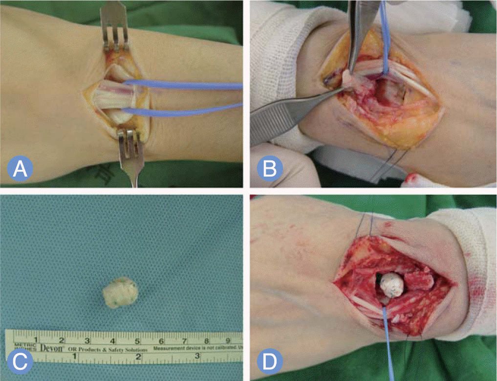 | Fig. 1.A 66-year-old female with Lichtman Class IIIB Kienbock’s disease. Third, fourth extensor compartment were found (A) and retracted to ulnar side (B). Tendon ball was made of Palmaris longus and plantaris tendons (C). After curettage of necrotic lunate bone tendon ball was inserted (D). |
 | Fig. 2.
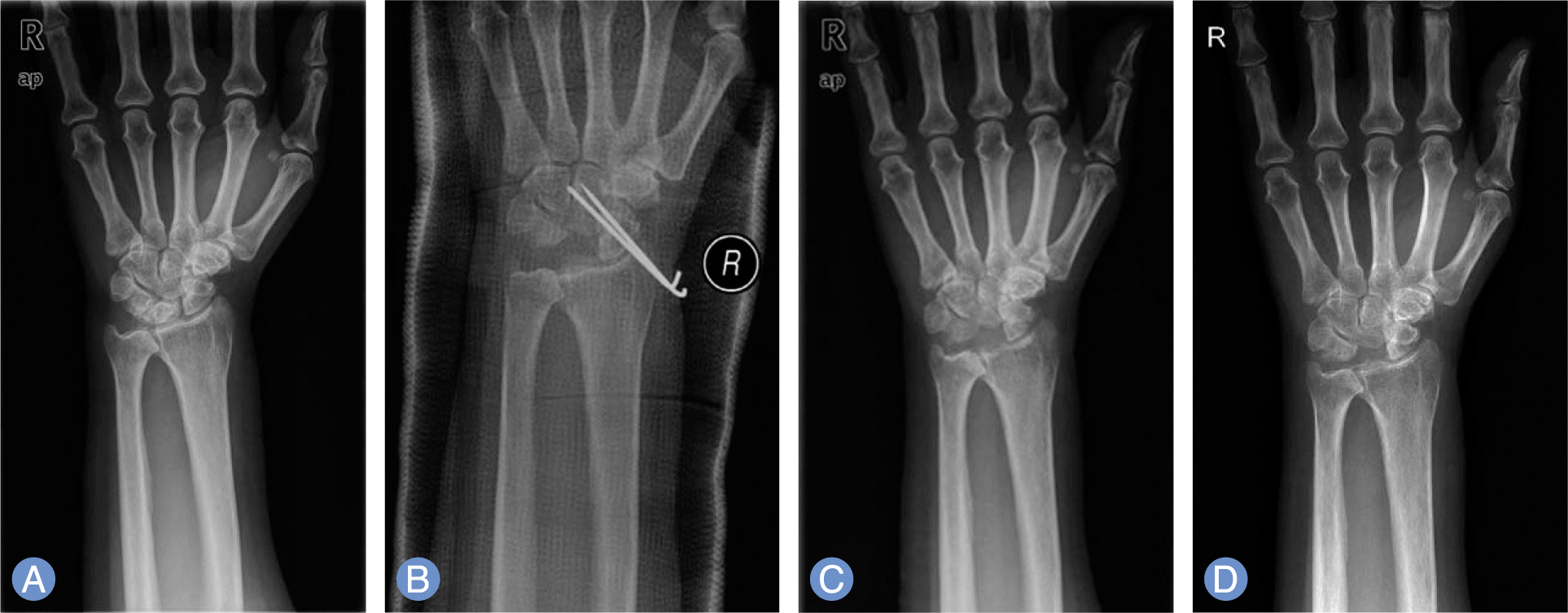
(A) Preoperative radiograph shows necrotic collapse of right lunate, Lichtman Class IIIB Kienbock’s disease. (B) Immediate postoperative radiograph shows excision of lunate, tendon transposition, temporary scaphocapitate fixation and thumb spica cast application. (C) 8 weeks postoperatively showing removal of K-wire and no residual bony fragment. (D) 45 months after surgery showing no further collapse or osteoarthritic change with calcification. |
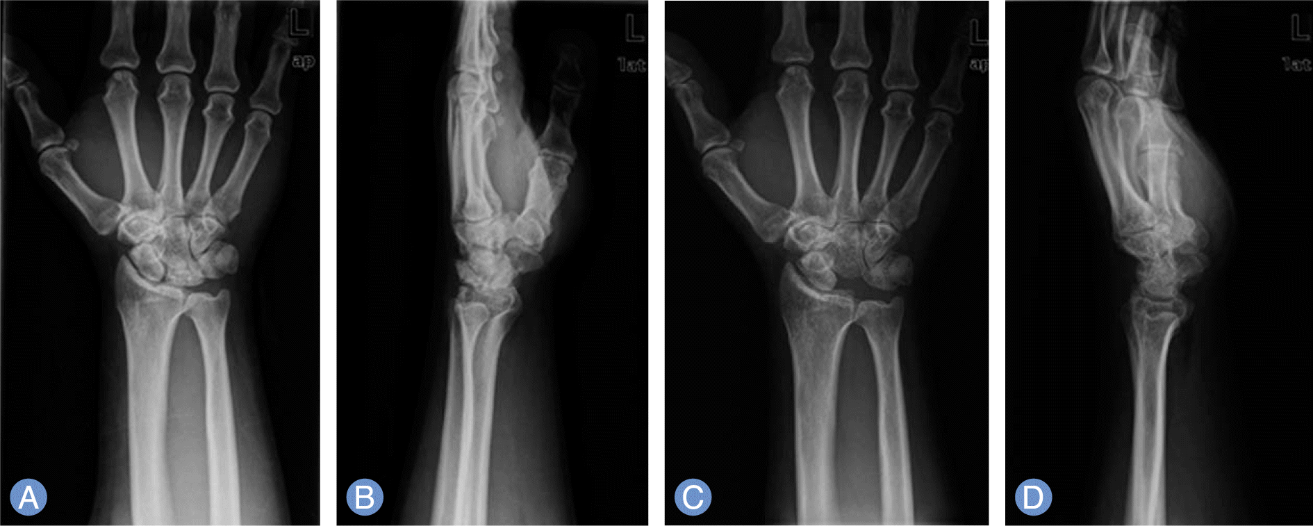 | Fig. 3.A 55-year-old male with Lichtman Class IIIB Kienbock’s disease (case No. 8). (A, B) Preoperative radiograph shows necrotic collapse of left lunate and ulnar negative varience. (C, D) 37 months after surgery showing no further collapse or osteoarthritic change with calcificaion. |
Table 1.
Clinical data of 10 patients operated for Kienböck’s disease
Table 2.
Postoperative and last follow-up change of carpal height ratio
 XML Download