

 PDF
PDF ePub
ePub Citation
 Print
Print


Abstract
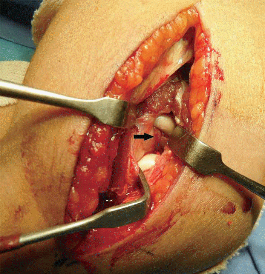
Traumatic dislocation of the radial head without fracture of the olecranon is very rare, especially in adults. We experienced a case of irreducible radial head dislocation with fracture without involvement of ulna. Open reduction and internal fixation was performed. During surgery, brachialis was interposed between capitellum and radial head, and also interposed between the fragments at the fracture site of the radial head. At 12 months after operation, the radial head was well reduced with normal rotation.
References
1. Watanabe K, Iwabu S, Hosoya T. Traumatic isolated anterior dislocation of the radial head in an adult: a case report. J Shoulder Elbow Surg. 2005; 14:554–6.

2. Sasaki K, Miura H, Iwamoto Y. Unusual anterior radial head dislocation associated with transposed biceps tendon: a case report. J Shoulder Elbow Surg. 2006; 15:e15–9.
3. Upasani VV, Hentzen ER, Meunier MJ, Abrams RA. Anteromedial radial head fracture-dislocation associated with a transposed biceps tendon: a case report. J Shoulder Elbow Surg. 2011; 20:e14–8.
4. Heo YM, Kim WS, Kim SH, Jeon TS, Kim SB, Oh BH. Isolated anterior dislocation of the radial head in adult: a case report. J Korean Shoulder Elbow Soc. 2007; 10:131–5.
5. Kong KC. Irreducible isolated dislocation of the radial head in a skeletally mature teenager. A case report. Arch Orthop Trauma Surg. 1993; 112:304–5.
6. Takami H, Takahashi S, Ando M. Irreducible isolated dislocation of the radial head. Clin Orthop Relat Res. 1997; (345):168–70.
7. Bonatus T, Chapman MW, Felix N. Traumatic anterior dislocation of the radial head in an adult. J Orthop Trauma. 1995; 9:441–4.
8. Baraza N, Saithna A, Krkovic MK. Acute persistent traumatic anterior dislocation of the fractured radial head: a case report and surgical technique. J Shoulder Elbow Surg. 2012; 21:e5–8.
Fig. 1.
(A) Anteroposterior and (B) lateral radiographs of the right elbow show anterior subluxation of the fractured radial head.
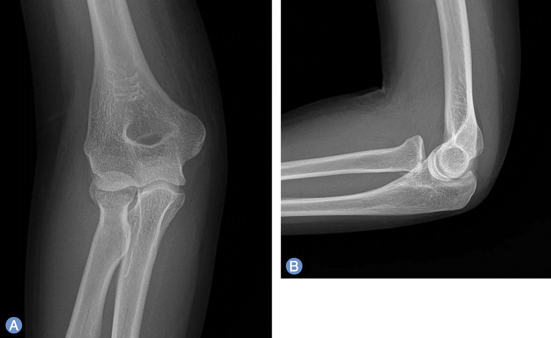
Fig. 2.
(A, B) Preoperative 3-dimensional computed tomography image shows radial head fracture and comminuted fragment, which displaced posteriorly and distally, and anterior subluxation of radial head.
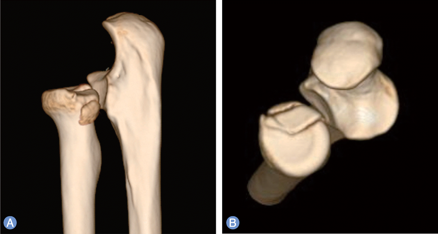
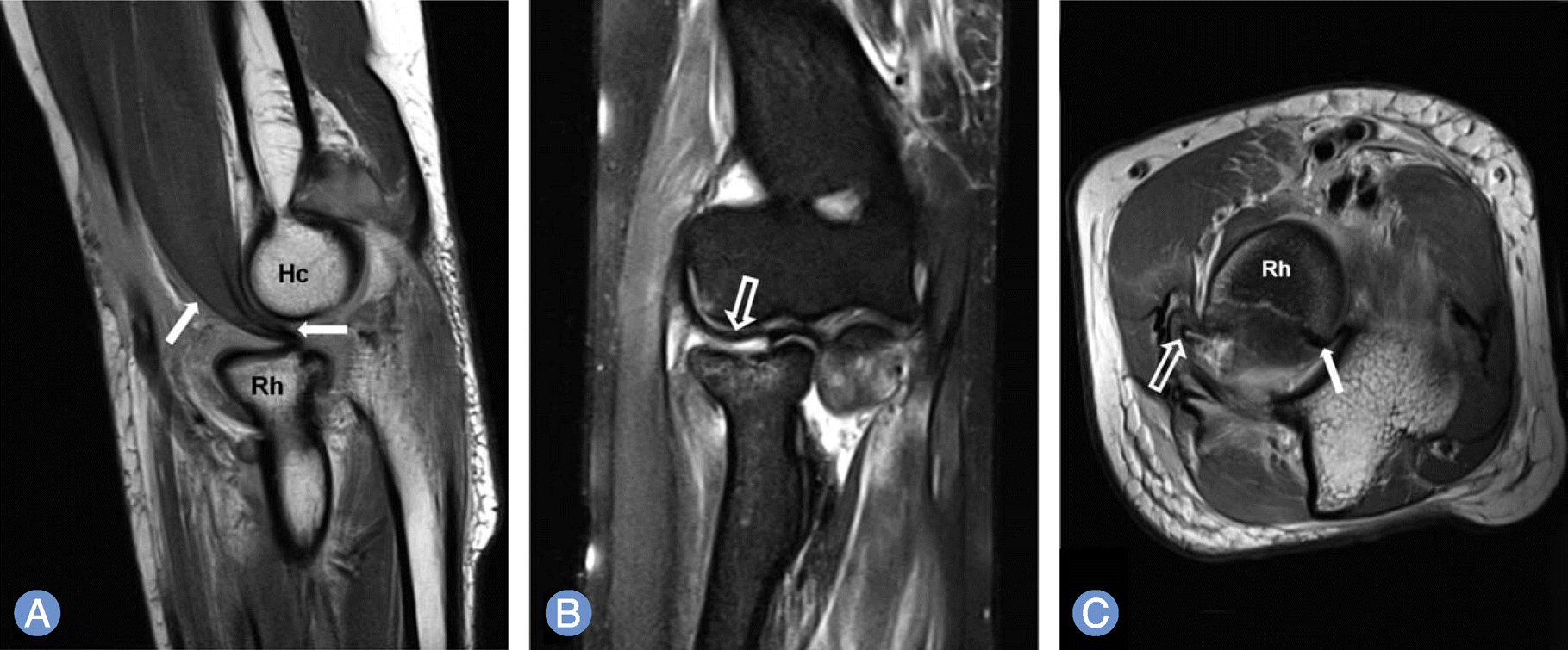
Fig. 3.
(A) Sagittal, proton density (PD)-weighted, fast spin echo (FSE) (repetition time [TR]/echo time [TE]: 3,310/24 msec) and (B) coronal fat suppression T2-weighted FSE (TR/TE: 4,000/52 msec) magnetic resonance (MR) images show the interposed brachialis muscle (arrows) and joint capsule (empty arrow) between anteriorly subluxed radial head (Rh) and the capitellum (Hc). (C) On axial PD-weighted FSE (TR/TE: 4,410/24 msec) MR images, the brachialis (arrow) and joint capsule (empty arrow) are also interposed between the fracture fragments of Rh.

 XML Download