

 PDF
PDF ePub
ePub Citation
 Print
Print


Abstract
Purpose:
Ulnar shortening osteotmy is a common operation for the treatment of ulnar impaction syndrome. The purpose of this study was to evaluate factors that may affect the occurrence of distal radioulnar joint (DRUJ) arthritis after ulnar shortening osteotomy.
Methods:
From September 2005 to August 2012, we performed 81 ulnar shortening osteotomies for ulnar impaction syndrome, and evaluated occurrence or deterioration of DRUJ arthritis in 58 patients with a minimum follow-up of 1 year. We analyzed potential factors that may affect the occurrence of DRUJ arthritis, such as, age, sex, hand dominance, pre- and postoperative ulnar variance, preexisting DRUJ arthritis, types of radial sigmoid notch, amount of ulnar shortening, and follow up period.
Results:
DRUJ arthritis occurred or deteriorated in 32 out of the 58 patients. Regression analysis indicated a significant correlation between the type of radial sigmoid notch (type 1) and DRUJ arthritis. Other factors were not found to be correlated with occurrence or deterioration of DRUJ arthritis.
Go to : 
References
1. Friedman SL, Palmer AK. The ulnar impaction syndrome. Hand Clin. 1991; 7:295–310.

2. Milch H. Colles’ fracture. Bull Hosp Joint Dis. 1950; 11:61–74.
3. Darrow JC Jr, Linscheid RL, Dobyns JH, Mann JM 3rd, Wood MB, Beckenbaugh RD. Distal ulnar recession for disorders of the distal radioulnar joint. J Hand Surg Am. 1985; 10:482–91.
4. Chun S, Palmer AK. The ulnar impaction syndrome: follow-up of ulnar shortening osteotomy. J Hand Surg Am. 1993; 18:46–53.
5. Palmer AK, Werner FW. Biomechanics of the distal radioulnar joint. Clin Orthop Relat Res. 1984; (187):26–35.
6. Kreder HJ, Hanel DP, McKee M, Jupiter J, McGillivary G, Swiontkowski MF. X-ray film measurements for healed distal radius fractures. J Hand Surg Am. 1996; 21:31–9.
7. Tolat AR, Stanley JK, Trail IA. A cadaveric study of the anatomy and stability of the distal radioulnar joint in the coronal and transverse planes. J Hand Surg Br. 1996; 21:587–94.
8. Kellgren JH, Lawrence JS. Radiological assessment of osteo-arthrosis. Ann Rheum Dis. 1957; 16:494–502.
9. Nakamura R, Tanaka Y, Imaeda T, Miura T. The influence of age and sex on ulnar variance. J Hand Surg Br. 1991; 16:84–8.
10. Sagerman SD, Zogby RG, Palmer AK, Werner FW, Fortino MD. Relative articular inclination of the distal radioulnar joint: a radiographic study. J Hand Surg Am. 1995; 20:597–601.
11. Baek GH, Lee HJ, Gong HS, et al. Long-term outcomes of ulnar shortening osteotomy for idiopathic ulnar impaction syndrome: at least 5-years follow-up. Clin Orthop Surg. 2011; 3:295–301.
12. Minami A, Kato H. Ulnar shortening for triangular fibrocartilage complex tears associated with ulnar positive variance. J Hand Surg Am. 1998; 23:904–8.
13. Koppel M, Hargreaves I, Herbert T. Ulnar shortening osteotomy for ulnar carpal instability and ulnar carpal impaction. J Hand Surg Eur Vol. 1997; 22:451–6.
14. Nishiwaki M, Nakamura T, Nakao Y, Nagura T, Toyama Y. Ulnar shortening effect on distal radioulnar joint stability: a biomechanical study. J Hand Surg Am. 2005; 30:719–26.
15. Kleinman WB, Graham TJ. The distal radioulnar joint capsule: clinical anatomy and role in posttraumatic limitation of forearm rotation. J Hand Surg Am. 1998; 23:588–99.
16. Schuind F, An KN, Berglund L, et al. The distal radioulnar ligaments: a biomechanical study. J Hand Surg Am. 1991; 16:1106–14.
17. Ward LD, Ambrose CG, Masson MV, Levaro F. The role of the distal radioulnar ligaments, interosseous membrane, and joint capsule in distal radioulnar joint stability. J Hand Surg Am. 2000; 25:341–51.
18. Baek GH, Chung MS, Lee YH, Gong HS, Lee S, Kim HH. Ulnar shortening osteotomy in idiopathic ulnar impaction syndrome. J Bone Joint Surg Am. 2005; 87:2649–54.
19. Miura T, Firoozbakhsh K, Cheema T, Moneim MS, Edmunds M, Meltzer S. Dynamic effects of joint-leveling procedure on pressure at the distal radioulnar joint. J Hand Surg Am. 2005; 30:711–8.
Go to :
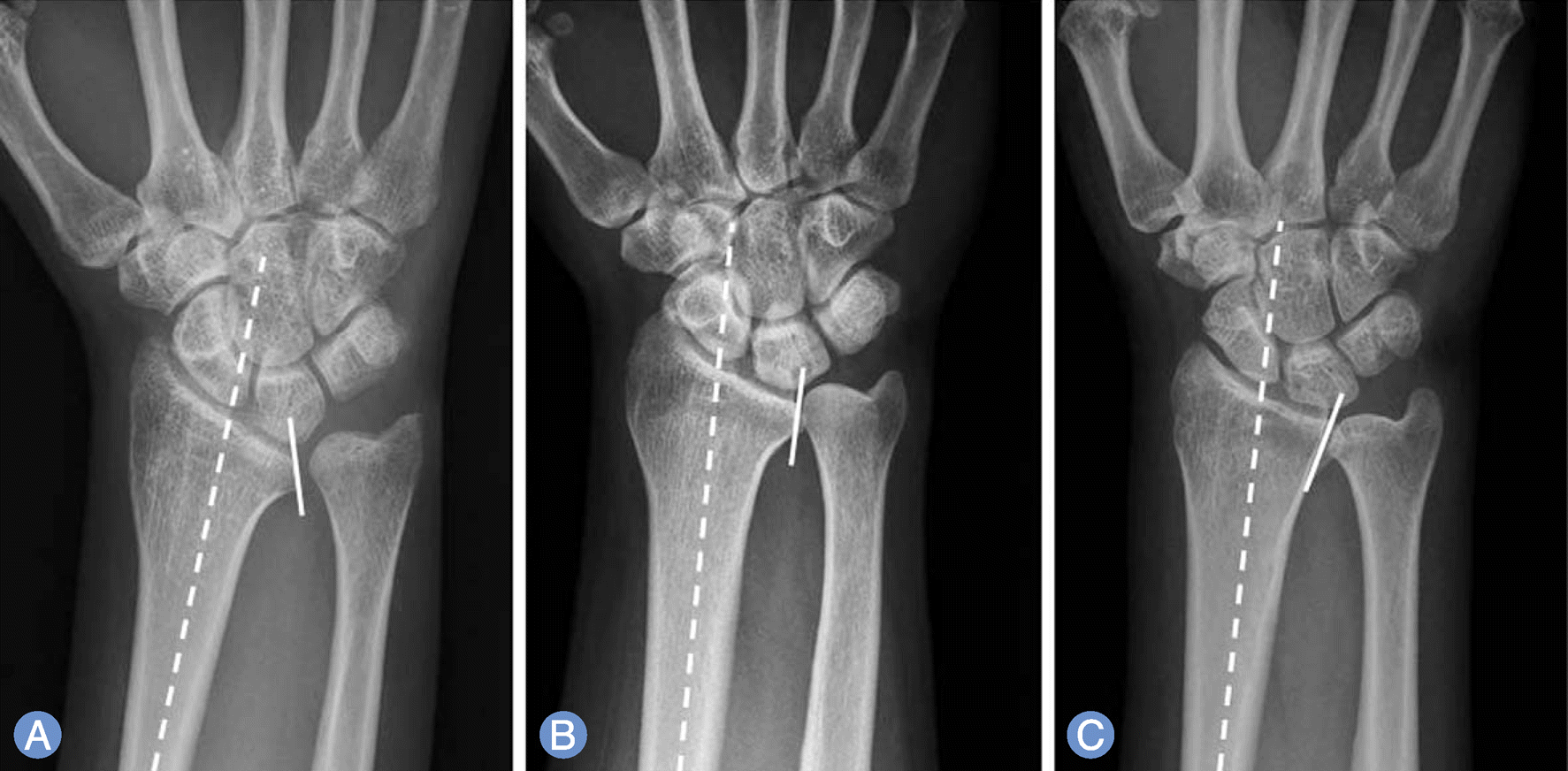 | Fig. 1.Types of the sigmoid notchs of distal radioulnar joints. (A) Type I is a hemispherical type that sigmoid notch is tilted to the ulnar side, more than 10 degrees. (B) Type II is a cylinderical type. Sigmoid notch is tilted less than 10 degrees to the radial or ulnar side. (C) Type III is a conical type that sigmoid notch tilted to radial side more than 10 degrees. |
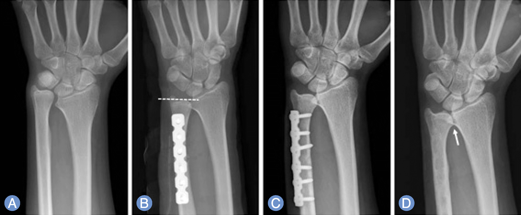 | Fig. 2.The patient with a complaining of right wrist pain for the past 1 year. (A) The radiograph showed 7 mm ulnar positive variance and sigmoid notch tilt to ulnar side (type II sigmoid notch). (B) Ulnar impaction syndrome was diagnosed and a ulnar shortening surgery was performed. The postoperative radiograph showed neutral ulnar variance. (C) Postoperative 14 months, there was no evidence of arthritic change. (D) Postoperative 27 months, there was little progression of arthritic change at distal radioulnar joint with sclerosis (white arrow). Visual analogue scale score was improved from 3 preoperatively to 0 at the last follow up and pain grade from 2 to 0 at the last follow-up. |
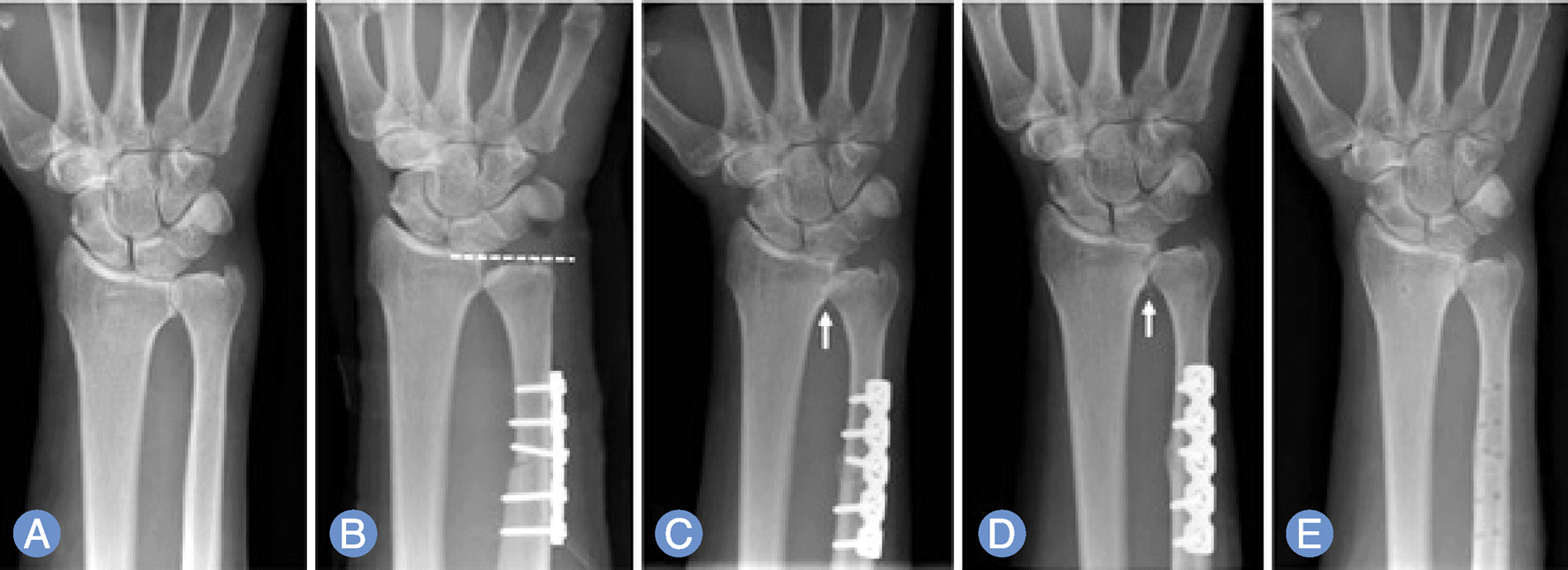 | Fig. 3.The patient has been complaining with a left wrist pain for five months. (A) The radiograph showed 5 mm ulnar positive variance and sigmoid notch tilt to ulnar side (type I). (B) A ulnar shortening surgery was performed. The postoperative radiograph showed 2 mm ulnar negative variance. (C) Postoperative 5 months, there was little osteophyte formation at distal radioulnar joint (white arrow). (D) Postoperative 9 months, progression of arthritic changes and more osteophyte formation were found (white arrow), which resulted in painful pronation/supination motion. (E) The osteophytes were excised. Visual analogue scale score was improved from 8 preoperatively to 2 at the last follow up and pain grade from 2 to 1 at postosteophyte-excision 18 months. |
Table 1.
Radiographic criteria for assessment of distal radioulnar joint arthritis
| Radiographic grade | Classification | Description |
|---|---|---|
| 0 | Normal | No features of arthritis |
| I | Doubtful | Sclerosis |
| II | Mild | Small*, single osteophyte |
| III | Moderate | Large†, multiple osteophyte |
| IV | Severe | Joint destruction |
Table 2.
Independent variable of distal radioulnar joint arthritis
Table 3.
Results of simple linear regression analysis for factors affecting on distal radioulnar joint arthritis
Table 4.
The outcomes of VAS score and pain grade
| Preoperative | At last follow-up | p-value | |
|---|---|---|---|
| VAS score | 6.29±2.38 | 1.63±1.65 | <0.05 |
| Pain grade* | 1.53±0.54 | 0.65 ±0.62 | <0.05 |
Table 5.
Comparison of VAS score and pain grade with/without DRUJ arthritis
 XML Download