Abstract
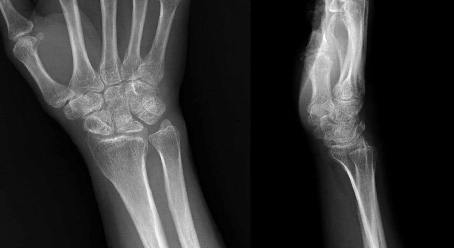
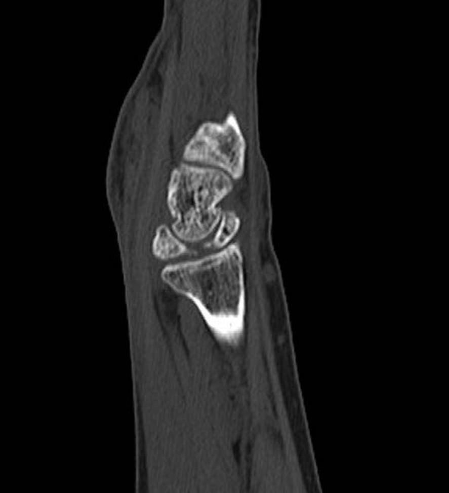
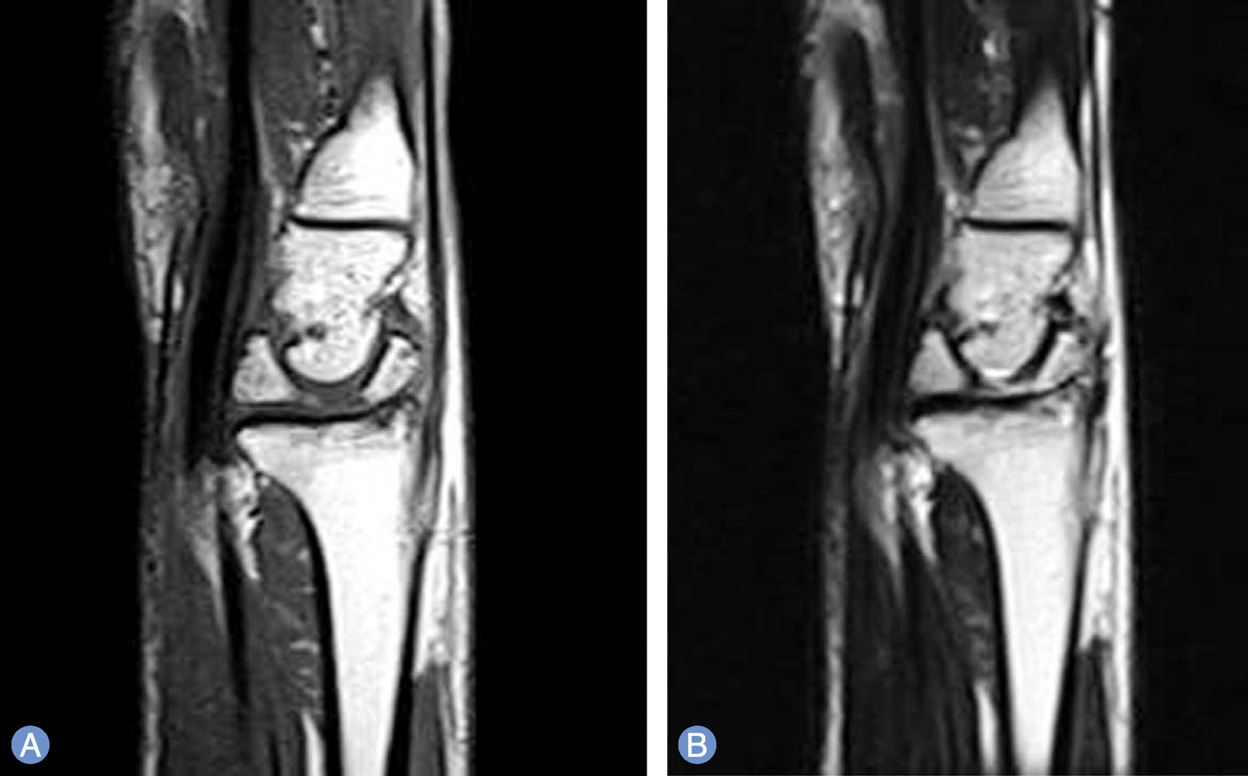
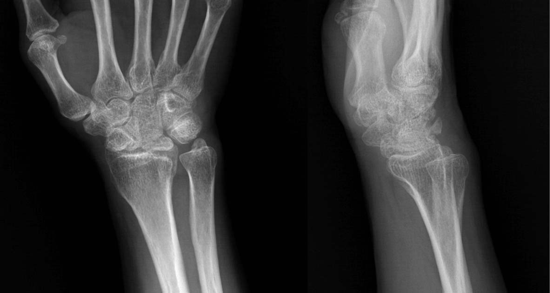
Isolate fracture of lunate is very rare. The authors reported a neglected fracture of lunate which was founded incidentally after the dorsal wall fracture of triquetrum. Pain reduction and improvement of range of motion was achieved after excising the dorsal fragment of lunate.
References
1. Lee SP, Jung JH, Kim JH, Lee KH, Chung US. Clinical results of perilunar fracture dislocation according to Mayfield stage and Herzberg stage. J Korean Soc Surg Hand. 2005; 10:59–65.
2. Teisen H, Hjarbaek J. Classification of fresh fractures of the lunate. J Hand Surg Br. 1988; 13:458–62.

3. Akahane M, Ono H, Sada M, Saitoh M. Bilateral bipartite lunate: a case report. J Hand Surg Am. 2002; 27:355–9.

4. Drez D Jr, Romero JR 3rd. Congenital bipartite carpal lunate: a case report. Am J Sports Med. 1978; 6:405–8.
5. Loh BW, Harvey J, Ek ET. Congenital bipartite lunate presenting as a misdiagnosed lunate fracture: a case report. J Med Case Rep. 2011; 5:102.

6. Luo J, Diao E. Kienbock's disease: an approach to treatment. Hand Clin. 2006; 22:465–73.
7. Galbraith PJ, Richardson ML. Fracture of the lunate: radiographic findings and case report. Case Rep Radiol. 2007; 2:13–6.

8. Hsu AR, Hsu PA. Unusual case of isolated lunate fracture without ligamentous injury. Orthopedics. 2011; 34:e785–9.


 PDF
PDF ePub
ePub Citation
 Print
Print


 XML Download