Abstract
Purpose:
The purpose of this study was to evaluate the clinical results of scaphoid nonunions treated with 1, 2-intercompartment supraretinacular artery (ICSRA) pedicled vascularized bone grafting (VBG) and headless compression screw fixation.
Methods:
Since August 1, 2005, 11 scaphoid nonunions with avascular necrosis or bone marrow edema of proximal fragments were managed with 1, 2-ICSRA pedicled VBG combined with headless compression screw fixation. The mean age was 37.1 years (range, 21-66 years). 8 patients had avascular necrosis (AVN) of proximal fragments and 3 patients had bone marrow edema in proximal fragments. Serial radiographic evaluations were performed in every 4-8 weeks for bone union and follow up computed tomography scanning were checked in 8 patients.
Results:
Bone unions were obtained in all 11 patients at 4.9 months (range, 3-9 months) after operation. At last follow up, the average range of motion was 82.5% and the grip power was 84.1% compared to the contralateral side. The mean New York Orthopaedic Hospital wrist score at last follow up was 83.2 (range, 58.1-93.3).
Go to : 
REFERENCES
1. Cooney WP 3rd, Dobyns JH, Linscheid RL. Nonunion of the scaphoid: analysis of the results from bone grafting. J Hand Surg Am. 1980; 5:343–54.

2. Cooney WP, Linscheid RL, Dobyns JH, Wood MB. Scaphoid nonunion: role of anterior interpositional bone grafts. J Hand Surg Am. 1988; 13:635–50.

3. Tomaino MM, King J, Pizillo M. Correction of lunate malalignment when bone grafting scaphoid nonunion with humpback deformity: rationale and results of a technique revisited. J Hand Surg Am. 2000; 25:322–9.

4. Green DP. The effect of avascular necrosis on Russe bone grafting for scaphoid nonunion. J Hand Surg Am. 1985; 10:597–605.

5. Pokorny JJ, Davids H, Moneim MS. Vascularized bone graft for scaphoid nonunion. Tech Hand Up Extrem Surg. 2003; 7:32–6.

6. Shin AY, Bishop AT. Pedicled vascularized bone grafts for disorders of the carpus: scaphoid nonunion and Kienbock's disease. J Am Acad Orthop Surg. 2002; 10:210–6.
7. Zaidemberg C, Siebert JW, Angrigiani C. A new vascularized bone graft for scaphoid nonunion. J Hand Surg Am. 1991; 16:474–8.

8. Nakamura R, Imaeda T, Horii E, Miura T, Hayakawa N. Analysis of scaphoid fracture displacement by three-dimensional computed tomography. J Hand Surg Am. 1991; 16:485–92.

9. McQueen MM, Gelbke MK, Wakefield A, Will EM, Gaebler C. Percutaneous screw fixation versus conservative treatment for fractures of the waist of the scaphoid: a prospective randomised study. J Bone Joint Surg Br. 2008; 90:66–71.
10. Gartland JJ Jr, Werley CW. Evaluation of healed Colles’ fractures. J Bone Joint Surg Am. 1951; 33:895–907.

11. Smith BS, Cooney WP. Revision of failed bone grafting for nonunion of the scaphoid. Treatment options and results. Clin Orthop Relat Res. 1996; (327):98–109.
12. Rajagopalan BM, Squire DS, Samuels LO. Results of Herbert-screw fixation with bone-grafting for the treatment of nonunion of the scaphoid. J Bone Joint Surg Am. 1999; 81:48–52.

13. Park MJ, Lee JS, Shin SK. Treatment of scaphoid nonunionusing a pedicled vascularized bone graft. J Korean Orthop Assoc. 2006; 41:871–6.

14. Jones DB Jr, Burger H, Bishop AT, Shin AY. Treatment of scaphoid waist nonunions with an avascular proximal pole and carpal collapse. A comparison of two vascularized bone grafts. J Bone Joint Surg Am. 2008; 90:2616–25.
15. Malizos KN, Dailiana ZH, Kirou M, Vragalas V, Xenakis TA, Soucacos PN. Longstanding nonunions of scaphoid fractures with bone loss: successful reconstruction with vascularized bone grafts. J Hand Surg Br. 2001; 26:330–4.

16. Doi K, Oda T, Soo-Heong T, Nanda V. Free vascularized bone graft for nonunion of the scaphoid. J Hand Surg Am. 2000; 25:507–19.

17. Boyer MI, von Schroeder HP, Axelrod TS. Scaphoid nonunion with avascular necrosis of the proximal pole. Treatment with a vascularized bone graft from the dorsum of the distal radius. J Hand Surg Br. 1998; 23:686–90.
18. Gunal I, Ozcelik A, Gokturk E, Ada S, Demirtas M. Correlation of magnetic resonance imaging and intraoperative punctate bleeding to assess the vascularity of scaphoid nonunion. Arch Orthop Trauma Surg. 1999; 119:285–7.
Go to : 
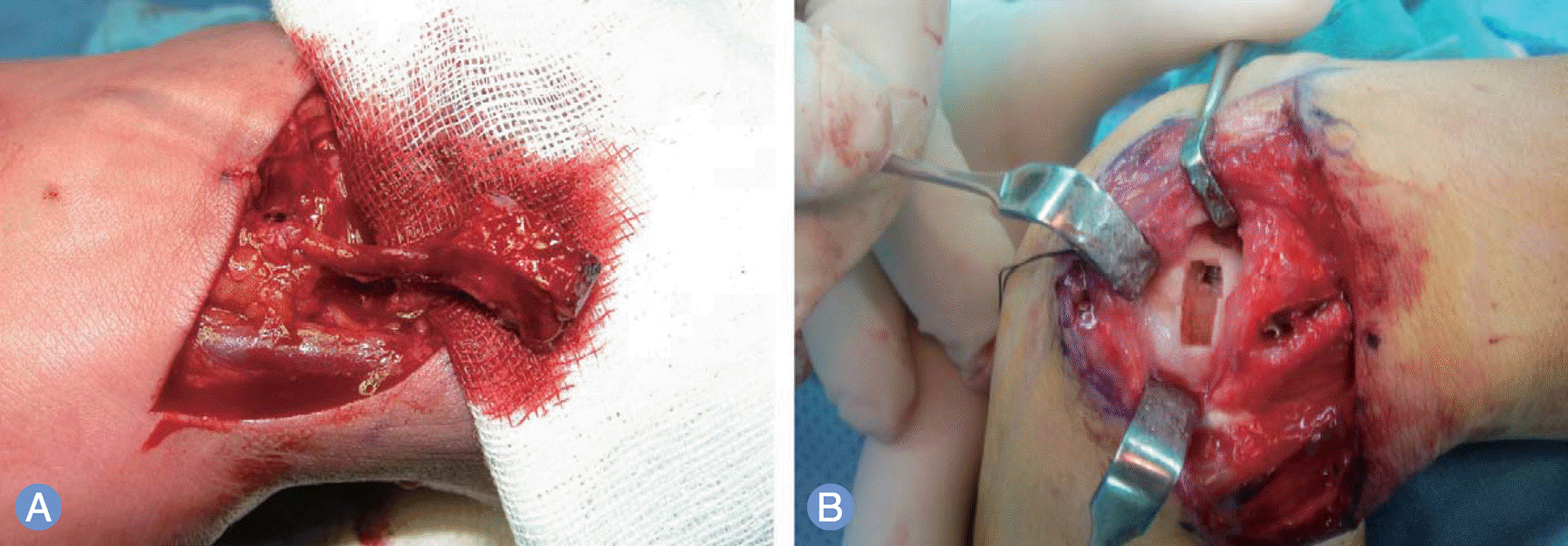
 | Fig. 1.Technique of 1, 2-intercompartment supraretinacular artery (ICSRA) pedicled vascularized bone graft using a dorsal approach for scaphoid nonunions. (A) The 1, 2-ICSRA pedicled vascularized bone graft was elevated from the dorsal surface of distal radius. The perfusion status of elevated bone graft was confirmed after tourniquet release. (B) After headless compression screw fixation, a slot for pedicled vascularized bone graft was prepared at dorsal surface of scaphoid with osteotome and curet. |
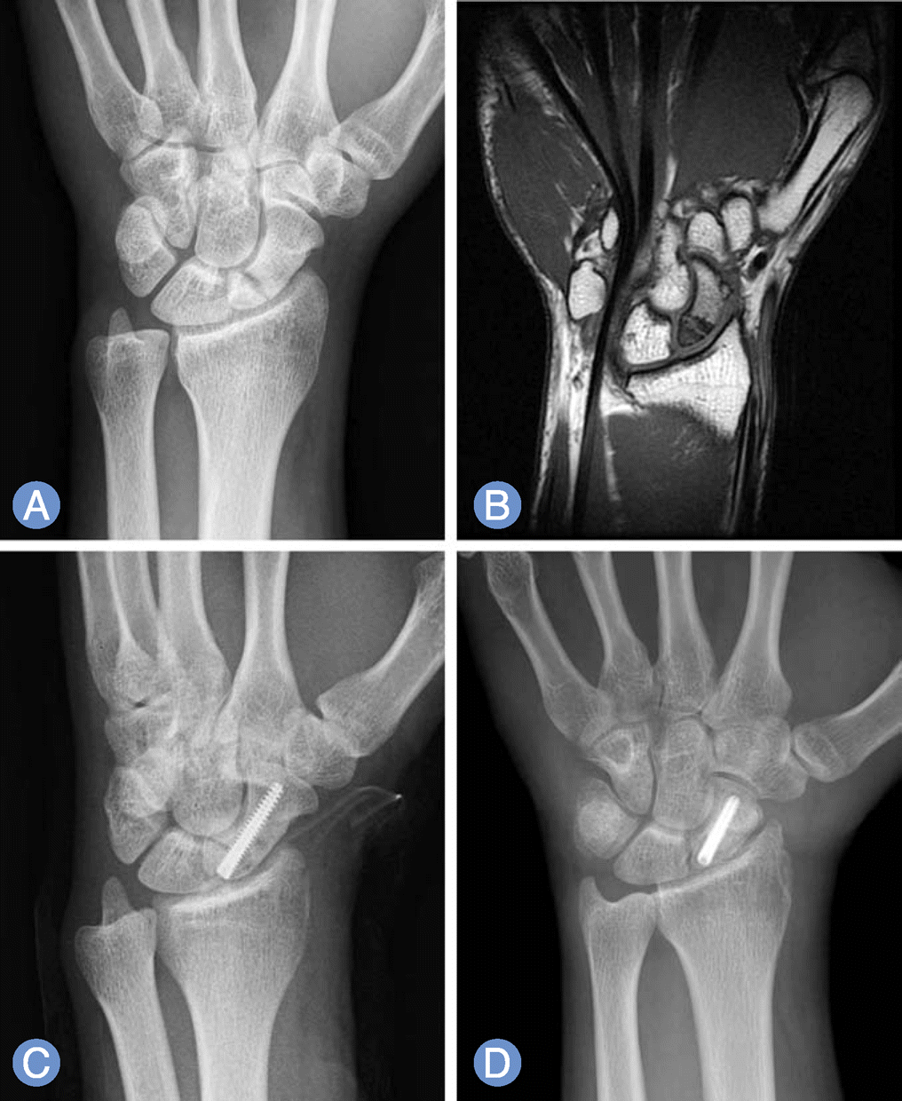
 | Fig. 2.The patient with a complaining of right wrist pain for 1 year. (A) The radiograph showed nonunion & sclerotic change of scaphoid proximal pole. (B) Magnetic resonance imaging showed avascular necrosis of proximal pole of scaphoid. (C) Headless compression screw fixation and 1, 2-intercompartment supraretinacular artery pedicled vascularized bone graft were performed. (D) At postoperative 3 months, complete bone union has progressed. |
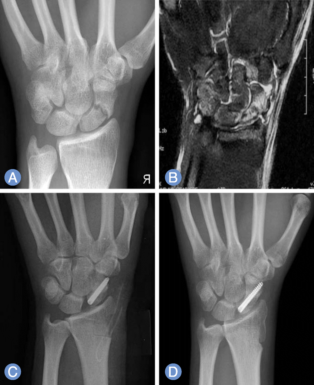
 | Fig. 3.The patient has been complaining with a right wrist pain for 1 year. (A) The radiograph showed scaphoid nonunion and sclerotic change. (B) Magnetic resonance imaging showed bone marrow edema of whole proximal fragment and large portion of distal fragment of scaphoid, which suggested precarious perfusion status. (C) 1, 2-intercompartment supraretinacular artery pedicled vascularized bone graft and headless compression screw fixation were performed. (D) At postoperative 12 months, computed tomography scan revealed progression of bone union and no more scaphoid collapse. |
Table 1.
Summary of cases (ROM, grip power, NYOH score, Green-O'Brien score and radiologic results)


 PDF
PDF ePub
ePub Citation
 Print
Print


 XML Download