

 PDF
PDF ePub
ePub Citation
 Print
Print


Abstract
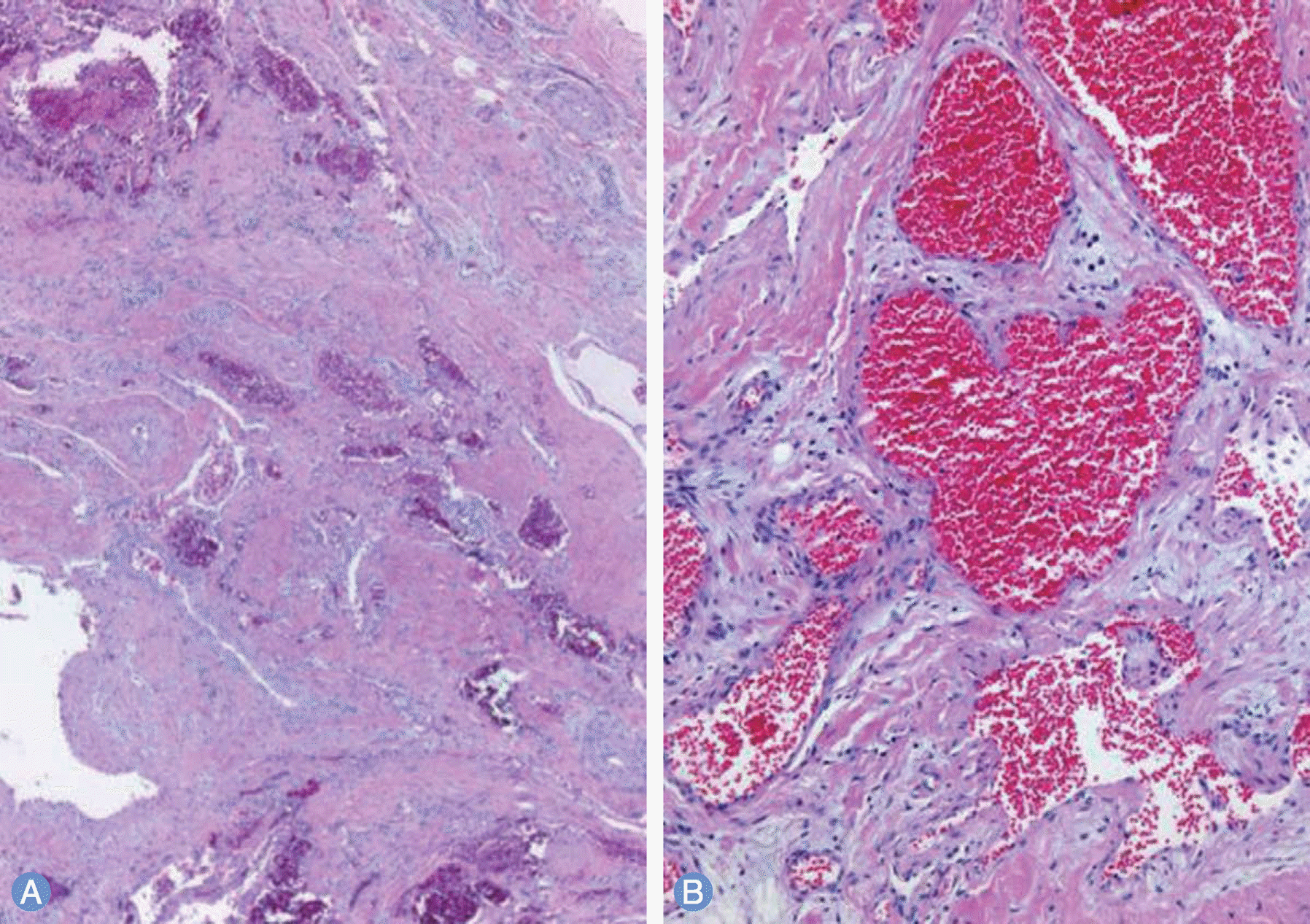
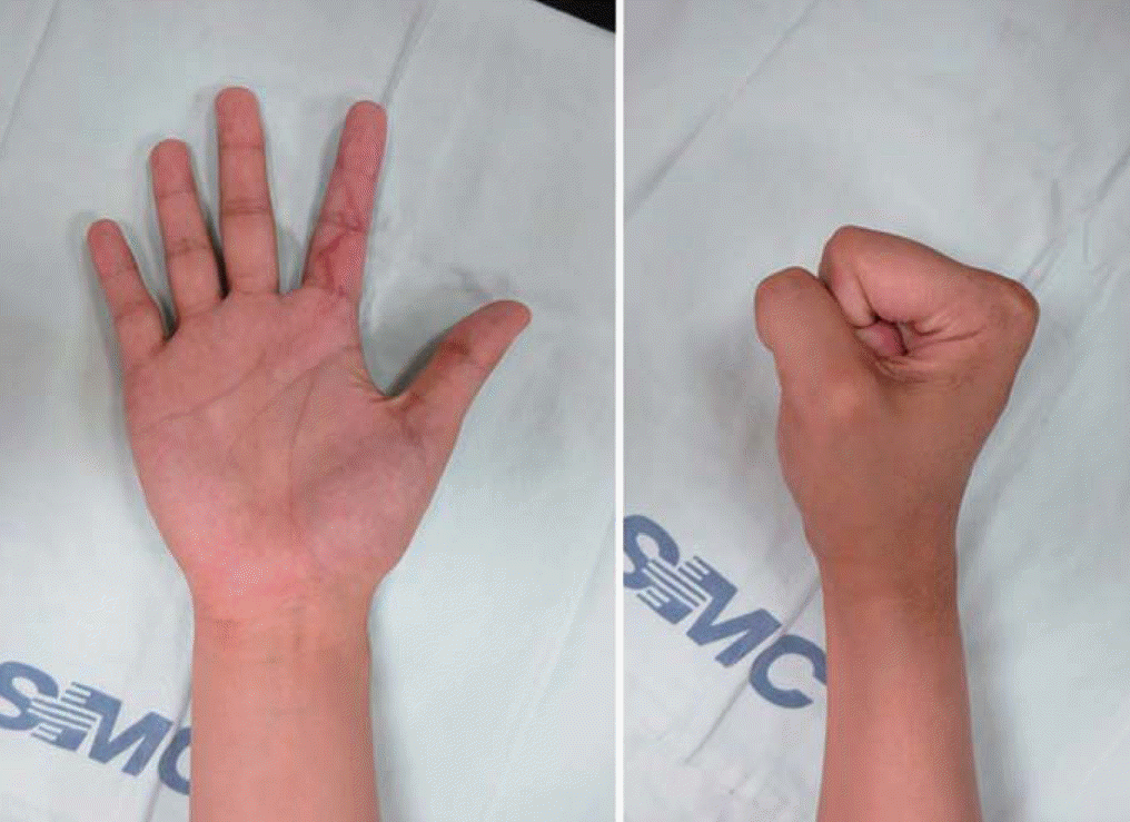
Hemangioma is a common tumor which can occur in any part of the body. It can develop in any area the hand. Hemangioma of the hand usually presents with swelling, pressure pain, accompanied by motion restriction. Rarely it has throbbing pain. We report the case of a 21-year-old woman who had a hemangioma of flexor digitorum superficialis tendon sheath in second finger with restriction of motion and were treated by surgical intervention.
Go to : 
REFERENCES
1. Devaney K, Vinh TN, Sweet DE. Synovial hemangioma: a report of 20 cases with differential diagnostic considerations. Hum Pathol. 1993; 24:737–45.

2. Chung DW, Lee JH, Lee JH. Intramuscular hemangioma of lumbrical muscle: a case report. J Korean Soc Surg Hand. 2002; 7:203–7.
3. Heo YM. Hemangioma of the flexor digitorum profundus of the hand: a case report. J Korean Soc Surg Hand. 2008; 13:142–5.
4. Lalonde D, Martin A. Epinephrine in local anesthesia in finger and hand surgery: the case for wide-awake anesthesia. J Am Acad Orthop Surg. 2013; 21:443–7.
5. Stack HG. Tumours of the Hand. Postgrad Med J. 1964; 40:290–8.
6. George R, Lee K. Synovial angioma of the FDP flexor sheath: a rare cause of carpal tunnel syndrome. Open Orthop J. 2013; 7:72–4.
7. Waddell GF. A hemangioma involving tendons. J Bone Joint Surg Br. 1967; 49:138–41.
8. Talwalkar S, Hayton M, Stilwell J, Temperley D, Freemont A. Tenosynovial haemangioma of the finger. Acta Orthop Belg. 2005; 71:618–21.
9. Rico AA, Holguin PH, Gonzalez IG, Coba JM. Flexor tendon synovial sheath haemangioma mimicking subacute tenosynovitis. J Hand Surg Br. 1994; 19:704–5.
10. Spinner M, Moon S, Young L. Recurrent cavernous haemangioma of the extensor tendons of the hand. Hand. 1983; 15:223–7.
Go to :
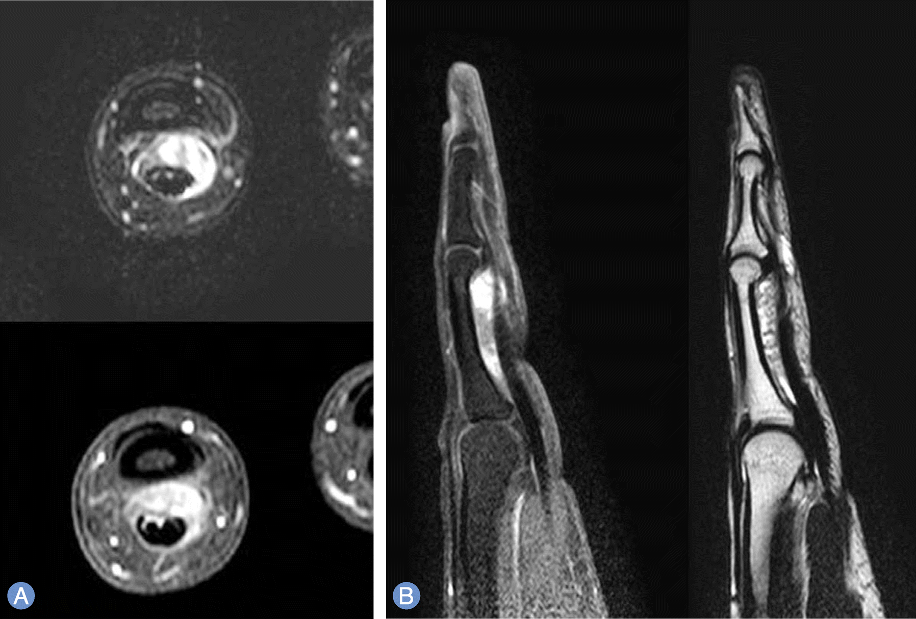 | Fig. 1.
(A) Axial view T1-weighted image shows that the signal of the lesion is equivalent to that of adjacent soft tissues. And T2-weighted image shows slightly ill-defined, lobulating slightly high signal intensity mass at the volar aspect of the proximal to middle phalanges level. (B) Sagittal view T1-weighted image shows ill-defined mixed high signal intensity nodule at the proximal phalanx level. And T2-weighted image shows that the high signal mass exist between bone and flexor tendons at the proximal phalanx level. |
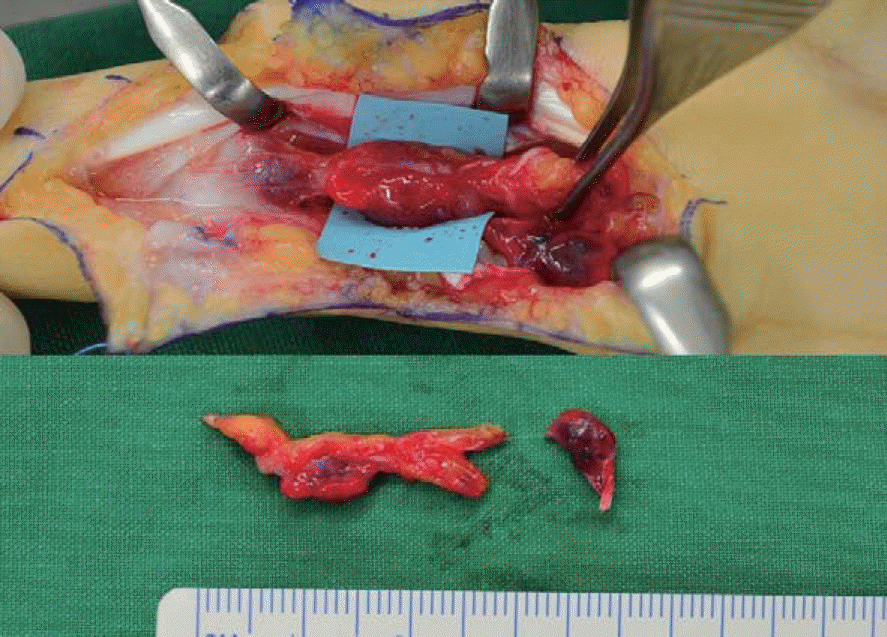 | Fig. 2.Intraoperative findings: the 2.9×0.6 cm sized fresh red tumor is identified between second proximal phalanx bone and flexor digitorum superficialis, and it is attached to tendon sheath and vincula longa distally. |
 XML Download