

 PDF
PDF ePub
ePub Citation
 Print
Print


Abstract
Purpose:
The purpose of this study was to present the results after functional reconstruction of the digits using palmaris longus tendocutaneous arterialized venous free flap in digits with compound defects.
Methods:
This study is based on 29 cases of palmaris longus tendocutaneous arterialized venous free flaps harvested from the ipsilateral wrist for the reconstruction of compound defect of the digits. Over the past 10 years, we performed in 17 cases of complex defects of extensor tendon on dorsum of the digits, 7 cases of collateral ligament of the proximal or distal interphalangeal joint and 5 cases of flexor tendon defect with soft tissue defect on the palmar aspect of the digits. We assessed survival rate of the flaps and functional recovery of the digits.
Results:
All free flaps completely survived except one with completele necrosis and another one with 50% necrosis. In cases of extensor tendon defect, the mean total active range of motion of the digits was 180°, in cases of flexor tendon reconstruction, it was 165°. In reconstruction of collateral ligament of interphalangeal joint of the thumb and digits, flexion and extension was within normal range and we got very good results without instability in all 7 cases.
REFERENCES
1. Nakayama Y, Soeda S, Kasai Y. Flaps nourished by arterial inflow through the venous system: an experimental investigation. Plast Reconstr Surg. 1981; 67:328–34.
2. Nishi G. Venous flaps for covering skin defects of the hand. J Reconstr Microsurg. 1994; 10:313–9.

3. Honda T, Nomura S, Yamauchi S, Shimamura K, Yoshimura M. The possible applications of a composite skin and subcutaneous vein graft in the replantation of amputated digits. Br J Plast Surg. 1984; 37:607–12.
4. Fasika OM, Stilwell JH. Arterialized venous flap for covering and revascularizing finger injury. Injury. 1993; 24:67–8.
5. Sandvall BK, Kuhlman-Wood K, Recor C, Friedrich JB. Flexor tendon repair, rehabilitation, and reconstruction. Plast Reconstr Surg. 2013; 132:1493–503.
6. Committee on rating of mental and physical impairment. The extremities and back. . Guides to the evaluation of permanent impairment. Chicago: American Medical Association;1971. p. 1–13.
7. Inoue G, Maeda N, Suzuki K. Resurfacing of skin defects of the hand using the arterialized venous flap. Br J Plast Surg. 1990; 43:135–9.
8. Woo SH, Seul JH. Pre-expanded arterialised venous free flaps for burn contracture of the cervicofacial region. Br J Plast Surg. 2001; 54:390–5.
9. Woo SH, Kim KC, Lee GJ, et al. A retrospective analysis of 154 arterialized venous flaps for hand reconstruction: an 11-year experience. Plast Reconstr Surg. 2007; 119:1823–38.
10. Kong BS, Kim YJ, Suh YS, Jawa A, Nazzal A, Lee SG. Finger soft tissue reconstruction using arterialized venous free flaps having 2 parallel veins. J Hand Surg Am. 2008; 33:1802–6.
11. Tsai TM, Matiko JD, Breidenbach W, Kutz JE. Venous flaps in digital revascularization and replantation. J Reconstr Microsurg. 1987; 3:113–9.
12. Yoshimura M, Shimada T, Imura S, Shimamura K, Yamauchi S. The venous skin graft method for repairing skin defects of the fingers. Plast Reconstr Surg. 1987; 79:243–50.
13. Woo SH, Jeong JH, Seul JH. Resurfacing relatively large skin defects of the hand using arterialized venous flaps. J Hand Surg Br. 1996; 21:222–9.
14. Abdul-Kader MH, Amin MA. Two-stage reconstruction for flexor tendon injuries in zone II using a silicone rod and pedicled sublimis tendon graft. Indian J Plast Surg. 2010; 43:14–20.
15. Wu WC, Wong TC, Yip TH. Chronic finger joint instability reconstructed with bone-ligament-bone graft from the iliac crest. J Hand Surg Br. 2004; 29:494–501.
16. Cho BC, Byun JS, Baik BS. Dorsalis pedis tendocutaneous delayed arterialized venous flap in hand reconstruction. Plast Reconstr Surg. 1999; 104:2138–44.
17. Sebastin SJ, Puhaindran ME, Lim AY, Lim IJ, Bee WH. The prevalence of absence of the palmaris longus: a study in a Chinese population and a review of the literature. J Hand Surg Br. 2005; 30:525–7.
18. Inoue G, Tamura Y. One-stage repair of both skin and tendon digital defects using the arterialized venous flap with palmaris longus tendon. J Reconstr Microsurg. 1991; 7:339–43.
19. Chen CL, Chiu HY, Lee JW, Yang JT. Arterialized tendocutaneous venous flap for dorsal finger reconstruction. Microsurgery. 1994; 15:886–90.
Fig. 1.
(A) Preoperative view of a compound defect on the volar aspect of the right index finger. (B) Flap design on the ipsilateral volar aspect of the distal forearm. (C, D) Dissected compound venous flap about 5.5×2 cm including the palmaris longus tendon and three veins (two blue arrows, efferent veins; red arrow, afferent vein; white arrow, palmaris longus tendon). (E) Repaired flexor tendon graft in zone I and II using a multiple ‘figure of 8’ technique. (F) Two dorsal veins were anastomosed. (G-J) Postoperative appearance 14 months later.
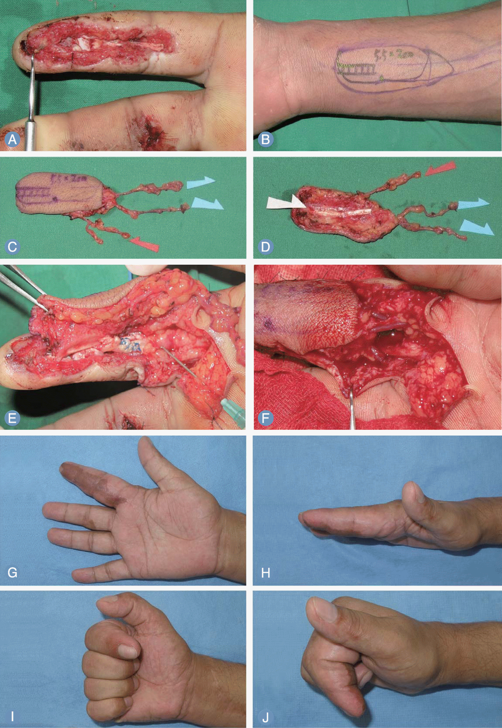
Fig. 2.
(A, B) Preoperative view of a compound defect on the dorsal aspect of the left long finger. (C, D) Flap design on the ipsilateral volar aspect of the distal forearm and dissected compound venous flap about 1.5×5 cm including the palmaris longus tendon and three veins (two blue arrows, efferent veins; red arrow, afferent vein). (E, F) Repaired extensor tendon and skin suture after microanastomosis of afferent and efferent veins. (G, H) Postoperative appearance of donor site and flap contour 12 months later. (I-L) Postoperative views of active motion of the long finger.
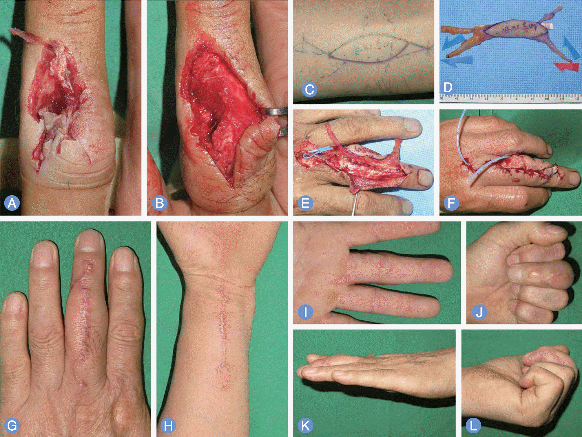
Fig. 3.
(A, B) Preoperative view of a compound defect on the ulanr side of distal interphalangeal joint of the left ring finger. (C, D) Flap design on the ipsilateral volar aspect of the distal forearm and dissected compound venous flap about 3×3 cm including the palmaris longus tendon and three veins (two blue arrows, efferent veins; red arrow, afferent vein). (E, F) Preoperative X-ray shows intraarticular fracture with difference of joint gap of distal interphalangeal joint. Postoperative radiography shows bone anchoring with micro screw with a little bulky soft tissue shadow. (G) Tendon anchoring with Mitek on distal and middle phalangeal bone. (H) Postoperative appearance of very strong stability against stress. (I-L) Postoperative appearance, 28 months later.

 XML Download