

 PDF
PDF ePub
ePub Citation
 Print
Print


Abstract
Purpose:
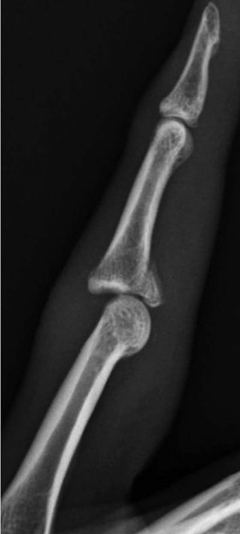
We report clinical and radiographic outcomes after internal fixation of intraarticular volar fractures of the middle phalanx base.
Methods:
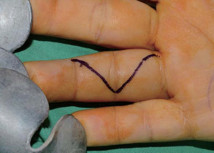
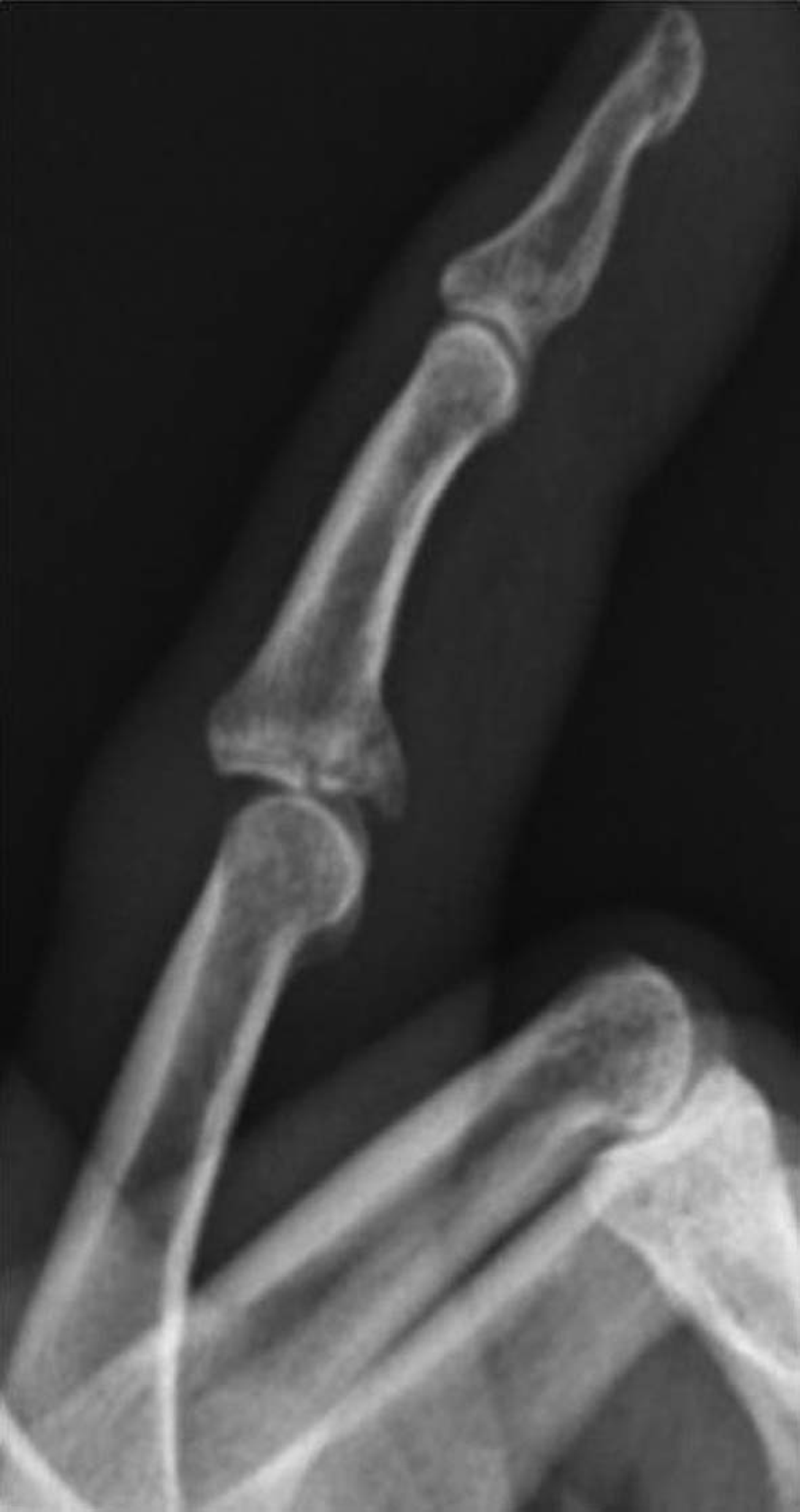
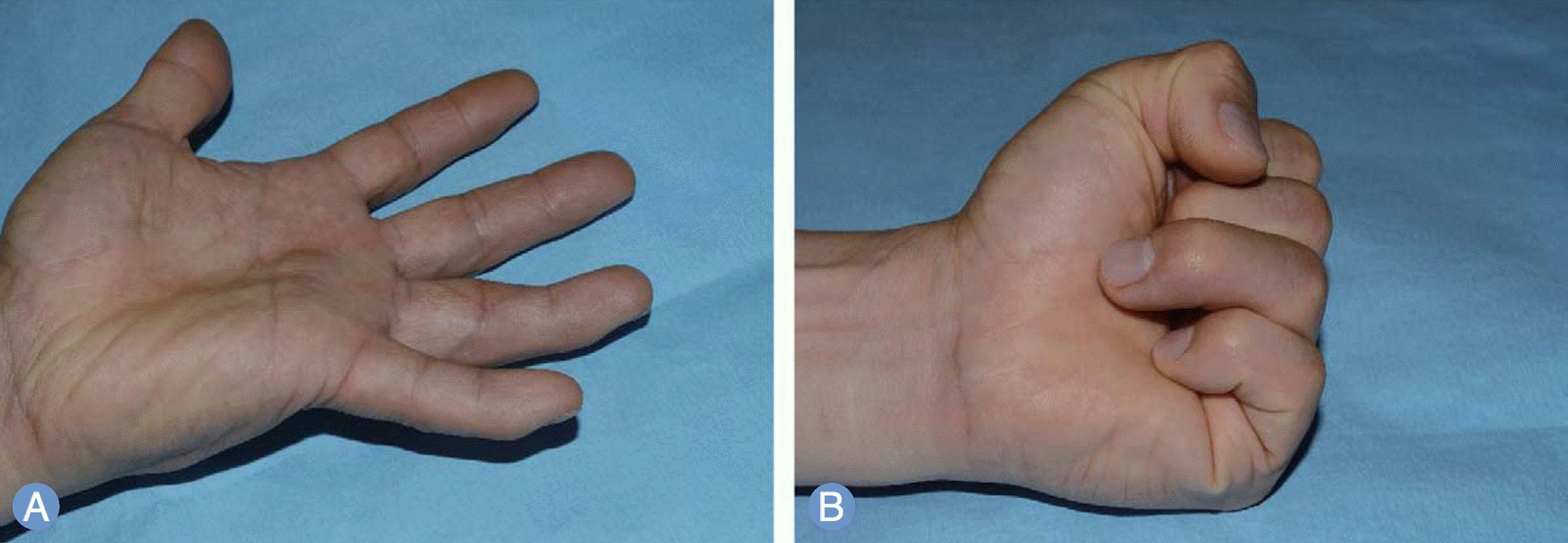
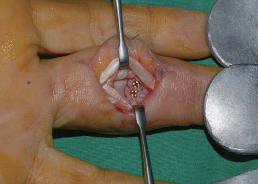
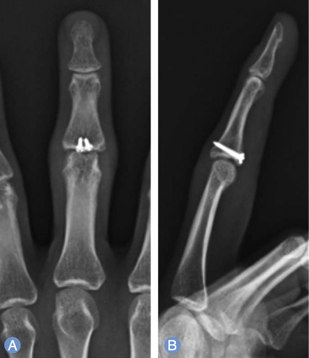
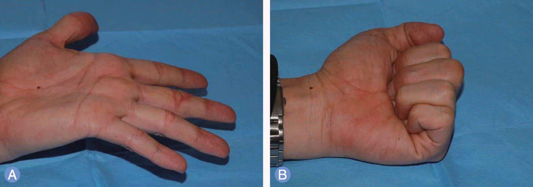
A retrospective review was conducted on 11 patients who had been treated with miniplate or screw for acute proximal interphalangeal joint fracture, after adjusting for the cases excluding severe comminuted or open fractures. The participants consisted of 9 males and 2 females, with average age of 43.7 years and with average follow-up period of 15.2 months. Bony union, change of articular side were examined through radiographs, and functional results were evaluated by means of the total active range of motion (TAM) and the disabilities of the arm, shoulder, and hand (DASH).
Results:
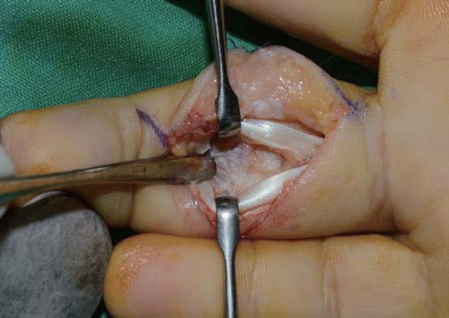
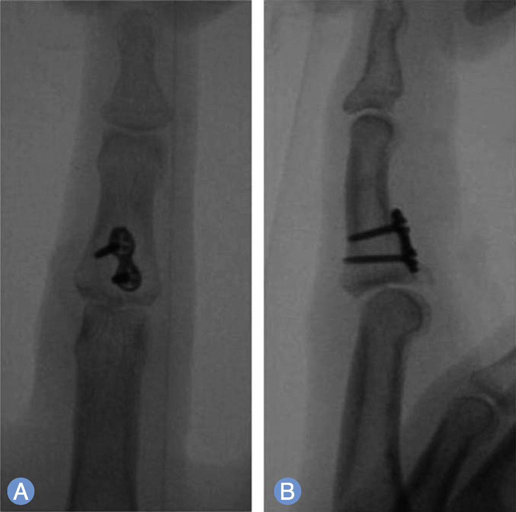
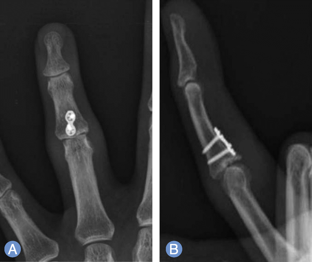
Of 11 cases of a finger fracture, 7 cases were fixed by miniplate with screws and 4 cases by screws alone. At the final follow-up retrospection, the average range of proximal interphalangeal joint motion was 95°, the average TAM was 243.2°, and the average DASH score was 7.4. Average 2.8 months was spent from the finger fracture to bony union. There was no case of degenerative change.
Go to : 
REFERENCES
1. Williams RM, Kiefhaber TR, Sommerkamp TG, Stern PJ. Treatment of unstable dorsal proximal interphalangeal fracture/dislocations using a hemi-hamate autograft. J Hand Surg Am. 2003; 28:856–65.

2. Ng CY, Watts AC. The use of non-vascularised osteo-chondral autograft for reconstruction of articular surfaces in the hand and wrist. J Bone Joint Surg Br. 2012; 94:1448–54.
3. Ikeda M, Kobayashi Y, Saito I, Ishii T, Shimizu A, Oka Y. Open reduction and internal fixation for dorsal fracture dislocations of the proximal interphalangeal joint using a miniplate. Tech Hand Up Extrem Surg. 2011; 15:219–24.
4. Cheah AE, Tan DM, Chong AK, Chew WY. Volar plating for unstable proximal interphalangeal joint dorsal fracture-dislocations. J Hand Surg Am. 2012; 37:28–33.
5. Waris E, Alanen V. Percutaneous, intramedullary fracture reduction and extension block pinning for dorsal proximal interphalangeal fracture-dislocations. J Hand Surg Am. 2010; 35:2046–52.
6. Suzuki Y, Matsunaga T, Sato S, Yokoi T. The pins and rubbers traction system for treatment of comminuted intraarticular fractures and fracture-dislocations in the hand. J Hand Surg Br. 1994; 19:98–107.
7. Eaton RG, Malerich MM. Volar plate arthroplasty of the proximal interphalangeal joint: a review of ten years’ experience. J Hand Surg Am. 1980; 5:260–8.
8. Seno N, Hashizume H, Inoue H, Imatani J, Morito Y. Fractures of the base of the middle phalanx of the finger. Classification, management and long-term results. J Bone Joint Surg Br. 1997; 79:758–63.
9. Mazhar FN. Proximal interphalangeal joint fracture dislocation. Shafa Ortho J. 2013; 1:29–33.
10. Wilson JN, Rowland SA. Fracture-dislocation of the proximal interphalangeal joint of the finger. J Bone Joint Surg Am. 1966; 48:493–502.
11. Cheah AE, Chong AK. Soft-tissue coverage of the hand. Curr Orthop Pract. 2012; 23:336–45.
Go to :
 XML Download