Abstract
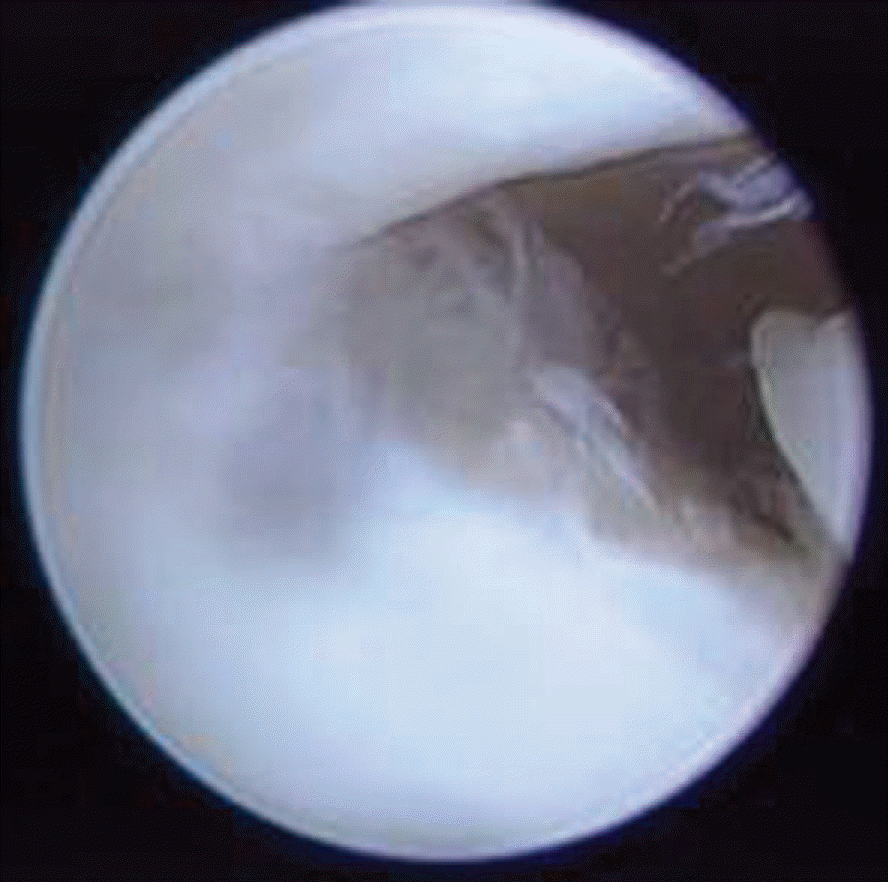
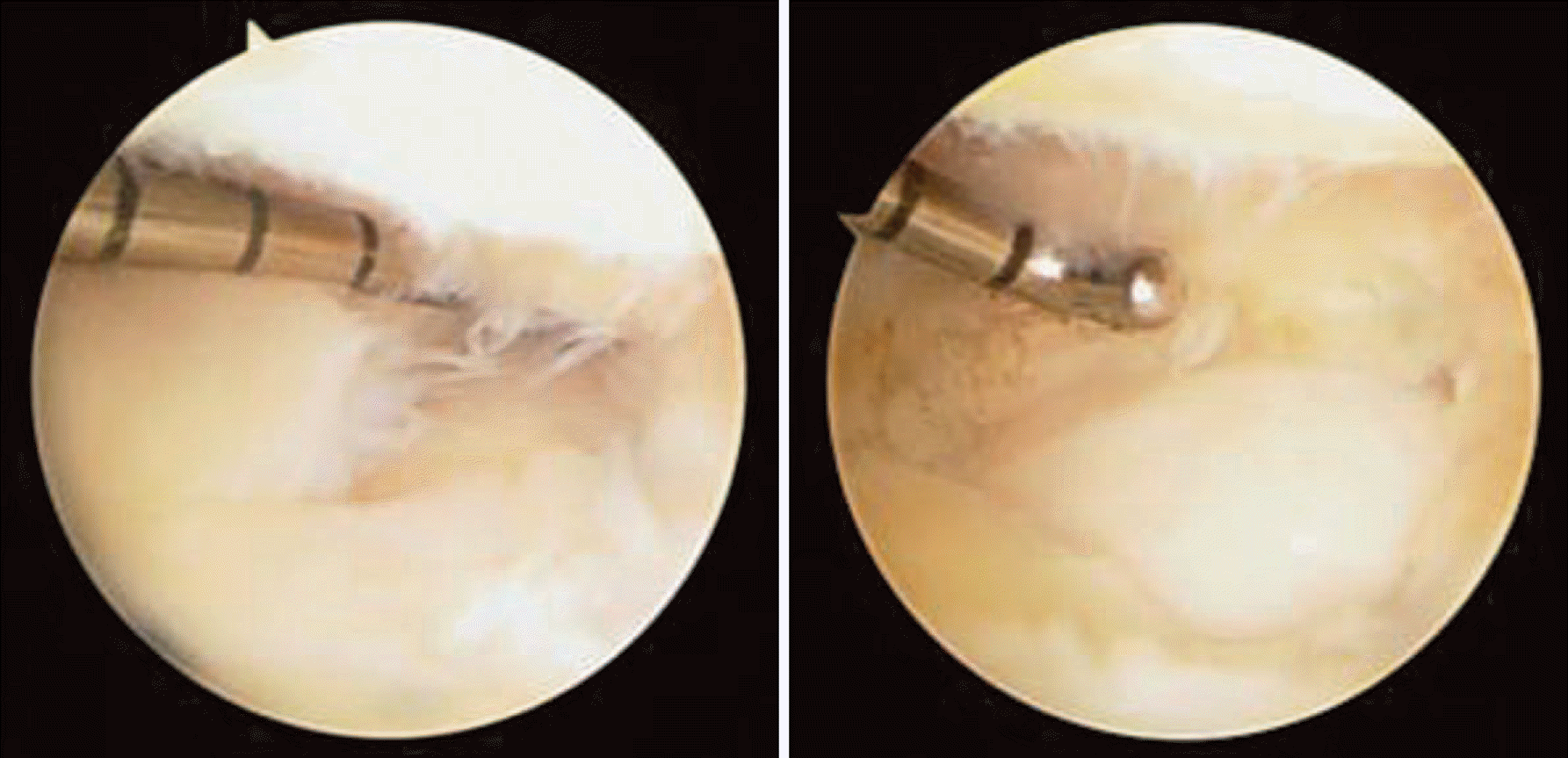
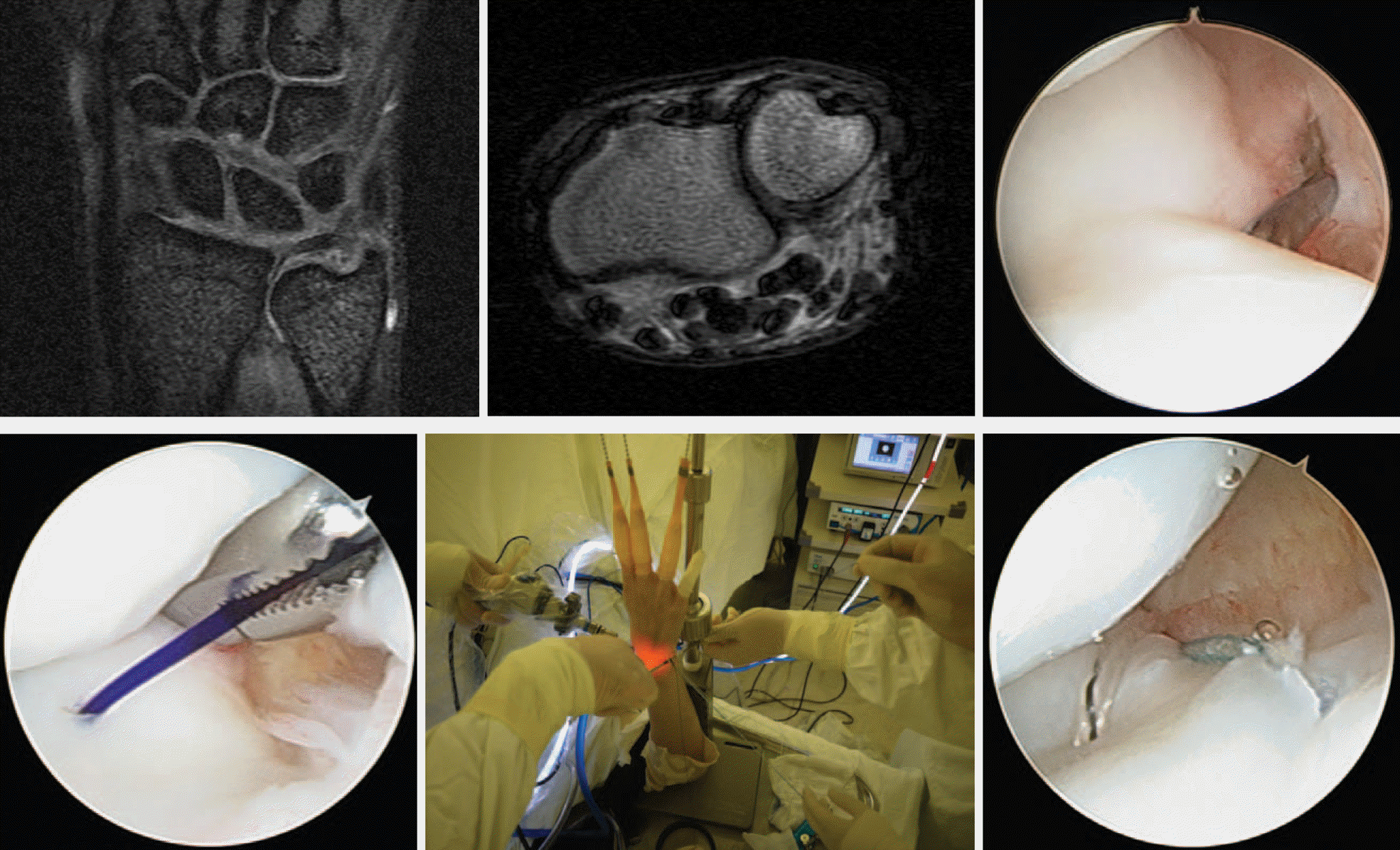
The Palmer class 1B triangular fibrocartilage complex injury has two entities: a lesion with stable distal radioulnar joint and a lesion with distal radioulnar joint instability. Arthroscopic debridement of fibrocartilage disk is used in Palmer class 1A lesion. The surgeon should remove the portion of the fibrocartilage tissue until a mechanically stable and smooth residual rim remains. Arthroscopic repair is used in Palmer class 1B or 1D lesion using meniscal repair sutures. Ulnar detachment that can produce distal radioulnar ligament instability can also be repaired using bone anchor or pull out suture. Old age as well as positive ulnar variance is poor prognostic factors.
Go to : 
REFERENCES
1. Adams BD. Distal radioulnar joint instability. Wolfe SW, Pederson WC, Hotchkiss RN, Kozin SH, editors. Green's operative hand surgery. 6th ed.Philadelphia, PA: Churchil Livinstone;2011. p. 523–60.

2. Henry MH. Management of acute triangular fbrocartilage complex injury of the wrist. J Am Acad Orthop Surg. 2008; 16:320–9.
3. Haugstvedt JR, Berger RA, Nakamura T, Neale P, Berglund L, An KN. Relative contributions of the ulnar attachments of the triangular fibrocartilage complex to the dynamic stability of the distal radioulnar joint. J Hand Surg Am. 2006; 31:445–51.

4. Tay SC, Tomita K, Berger RA. The “ulnar fovea sign” for defining ulnar wrist pain: an analysis of sensitivity and specificity. J Hand Surg Am. 2007; 32:438–44.

5. Tanaka T, Yoshioka H, Ueno T, Shindo M, Ochiai N. Comparison between high-resolution MRI with a microscopy coil and arthroscopy in triangular fibrocartilage complex injury. J Hand Surg Am. 2006; 31:1308–14.

6. Atzei A, Rizzo A, Luchetti R, Fairplay T. Arthroscopic foveal repair of triangular fibrocartilage complex peripheral lesion with distal radioulnar joint instability. Tech Hand Up Extrem Surg. 2008; 12:226–35.

7. Wysocki RW, Richard MJ, Crowe MM, Leversedge FJ, Ruch DS. Arthroscopic treatment of peripheral triangular fibrocartilage complex tears with the deep fibers intact. J Hand Surg Am. 2012; 37:509–16.

8. Corso SJ, Savoie FH, Geissler WB, Whipple TL, Jiminez W, Jenkins N. Arthroscopic repair of peripheral avulsions of the triangular fibrocartilage complex of the wrist: a multicenter study. Arthroscopy. 1997; 13:78–84.

9. Estrella EP, Hung LK, Ho PC, Tse WL. Arthroscopic repair of triangular fibrocartilage complex tears. Arthroscopy. 2007; 23:729–37.

10. Ruch DS, Papadonikolakis A. Arthroscopically assisted repair of peripheral triangular fibrocartilage complex tears: factors affecting outcome. Arthroscopy. 2005; 21:1126–30.

11. Wolf MB, Kroeber MW, Reiter A, et al. Ulnar shortening after TFCC suture repair of Palmer type 1B lesions. Arch Orthop Trauma Surg. 2010; 130:301–6.

12. Anderson ML, Larson AN, Moran SL, Cooney WP, Amrami KK, Berger RA. Clinical comparison of arthroscopic versus open repair of triangular fibrocartilage complex tears. J Hand Surg Am. 2008; 33:675–82.

13. Miwa H, Hashizume H, Fujiwara K, Nishida K, Inoue H. Arthroscopic surgery for traumatic triangular fibrocartilage complex injury. J Orthop Sci. 2004; 9:354–9.

Go to : 


 PDF
PDF ePub
ePub Citation
 Print
Print


 XML Download