Abstract
Triggering at the wrist during active flexion and extension of the fingers or wrists is very rare. It is caused by tumors, inflammation, and anomalous muscle belly. We report two cases of trigger wrist caused by synovial hypertrophy and fibroma of flexor tendon.
REFERENCES
1. Parmaksizoglu F, Beyzadeoglu T, Yildirim S. Haemangioma originating from a tendon sheath as an unusual cause of trigger wrist: case report. Handchir Mikrochir Plast Chir. 2003; 35:64–5.

2. Smith RD, O'Leary ST, McCullough CJ. Trigger wrist and flexor tenosynovitis. J Hand Surg Br. 1998; 23:813–4.

3. Sonoda H, Takasita M, Taira H, Higashi T, Tsumura H. Carpal tunnel syndrome and trigger wrist caused by a lipoma arising from flexor tenosynovium: a case report. J Hand Surg Am. 2002; 27:1056–8.

4. Suematsu N, Hirayama T, Takemitsu Y. Trigger wrist caused by a giant cell tumour of tendon sheath. J Hand Surg Br. 1985; 10:121–3.

7. Yamazaki H, Uchiyama S, Kato H. Snapping wrist caused by tenosynovitis of the extensor carpi radialis longus tendon subsequent to subcutaneous muscle rupture in the forearm: case report. J Hand Surg Am. 2010; 35:1964–7.

8. Desai SS, Pearlman HS, Patel MR. Clicking at the wrist due to fibroma in an anomalous lumbrical muscle: a case report and review of literature. J Hand Surg Am. 1986; 11:512–4.

9. Kernohan JG, Benjamin A, Simpson D. Trigger wrist due to anomalous flexor digitorum profundus muscle in association with fibroma of tendon sheath. Hand. 1982; 14:59–60.

11. Zachee B, DeSmet L, Fabry G. A snapping wrist due to a loose body. Arthroscopic diagnosis and treatment. Arthroscopy. 1993; 9:117–8.

12. Weeks PM, Young VL, Gilula LA. A cause of painful clicking wrist: a case report. J Hand Surg Am. 1979; 4:522–5.

13. Eibel P. Trigger Wrist with Intermittent Carpal Tunnel Syndrome: A Hitherto Undescribed Entity with Report of a Case. Can Med Assoc J. 1961; 84:602–5.
14. Carvell JE, Mowat AG, Fuller DJ. Trigger wrist phenomenon in rheumatoid arthritis. Hand. 1983; 15:77–81.

15. Minami A, Ogino T. Trigger wrist caused by a partial laceration of the flexor superficialis tendon of the ring finger. J Hand Surg Br. 1986; 11:457–9.

Fig. 1.
(A) Sagittal T2 magnetic resonance images shows nodular mass (arrows) surrounded by the flexor tendons at the just distal level to the transverse carpal ligament. The mass shows heterogeneously increased signal intensity on T2-weighted image. (B) On the contrast enhanced axial image, the lesion shows eccentric nodular and peripheral enhancement within the mass (arrows). Flattening and weak enhancement of the median nerve suggest entrapment neuropathy (arrowhead).
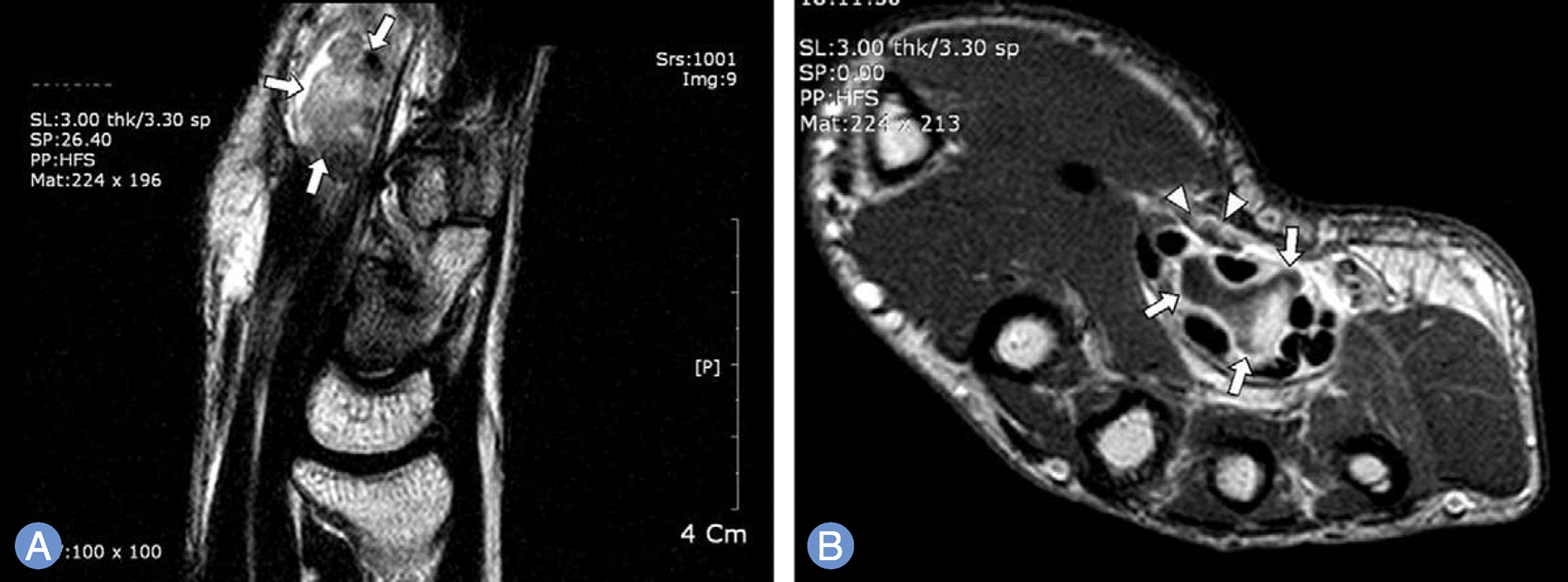
Fig. 2.
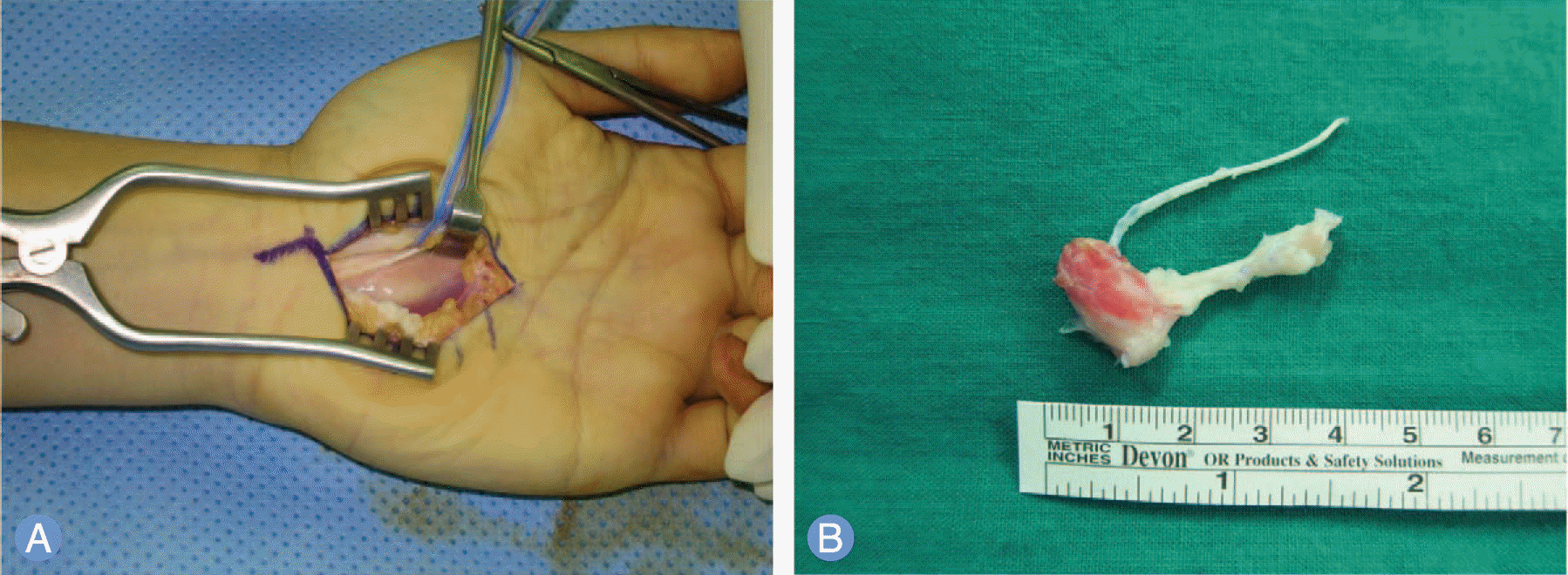
(A) Irregularly shaped nodular and streaky portion of the mass was freely separated from the tendon. However the proximal end of the mass was fixed to the tendon at the level of proximal entry of carpal tunnel. (B) A photograph taken just after the mass removal. There was no noticeable abnormality of tendon or tendon sheath. Note the separate sheath that encapsulated the mass. (C) A photograph of the mass.

Fig. 3.
A picture of low power microscopic examination (×100) with H&E staining shows portions of muscle bellies (arrows) and surrounding synovial, capillary and fibrous overgrowth. There is no inflammatory cell infiltration except scanty macrophages.
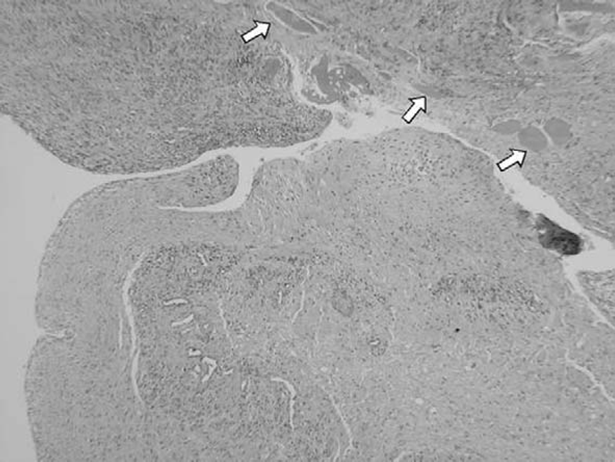
Fig. 4.
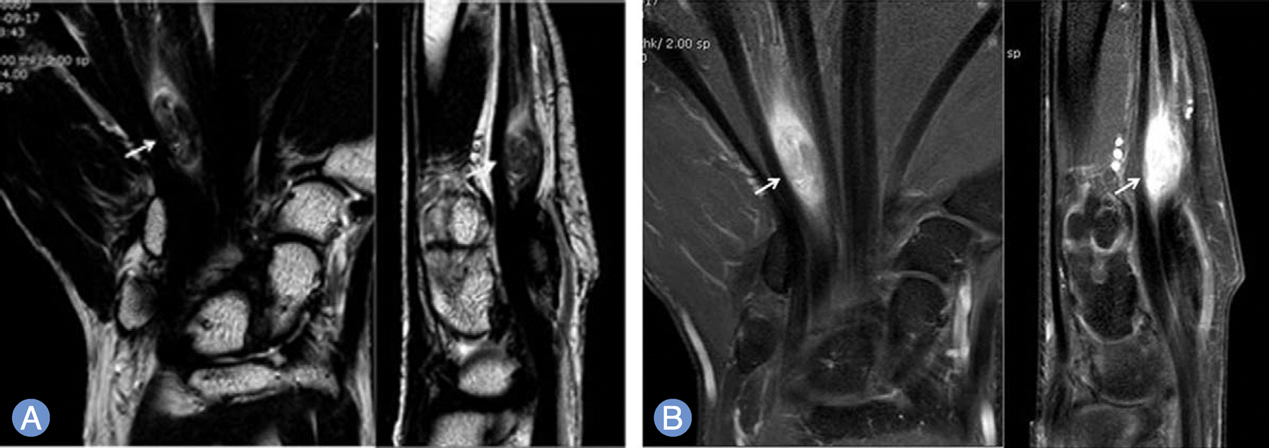
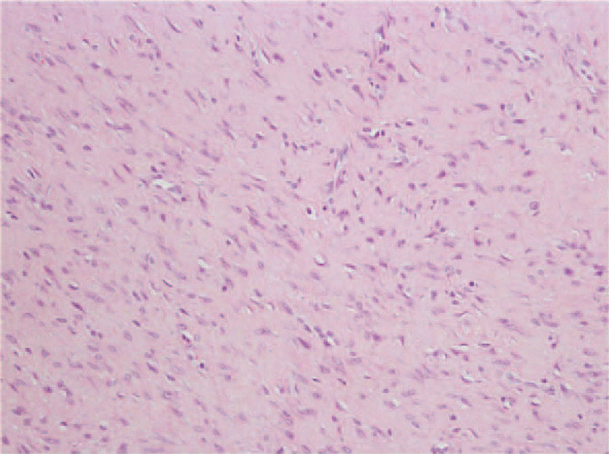
(A) T2-weighted magnetic resonance images show well-defined oval mass (arrows) with low heteogenous signal intensity between flexor tendon underneath transverse carpal ligament. (B) Enhanced images show heterogenous enhance-


 PDF
PDF ePub
ePub Citation
 Print
Print


 XML Download