Abstract
Purpose:
To evaluate the clinical and radiographic outcomes of scaphoid nonunion patients who had treated by open reduction and internal fixation with Herbert screw through dorsal approach.
Methods:
We reviewed prospectively a series of 102 consecutive patients with scaphoid nonunion (Mack-Lichtman stage I, II, III). All patients were managed with open reduction with dorsal approach and internal fixation with a Herbert screw and additional K-wires. Exclusion criteria included conservative treatment, percutaneous fixation, scaphoid nonunion advanced collapse wrist. There were 94 male and 8 female with an average age of 28 years (range, 13–65 years). The mean follow period was 35 months (range, 12–96 months). Postoperative radiographs were reviewed to assess the fracture union, carpal alignment, and screw position. Functional results were evaluated by modified Mayo wrist score.
Results:
Ninety-eight of 102 patients (96.1%) showed radiographic union at an average time of 12.7 weeks. Modified Mayo wrist score was 87.5 points in an average. Ninety-two of 102 patinets (91.3%) showed more than good results. There was no major complications. There was no statistically significant difference between the preoperative and postoperative radiolunate angle, scapholunate angle, or height to length scaphoid ratio.
Go to : 
REFERENCES
1. Chan KW, McAdams TR. Central screw placement in percutaneous screw scaphoid fixation: a cadaveric comparison of proximal and distal techniques. J Hand Surg Am. 2004; 29:74–9.

2. Braga-Silva J, Peruchi FM, Moschen GM, Gehlen D, Padoin AV. A comparison of the use of distal radius vascularised bone graft and non-vascularised iliac crest bone graft in the treatment of non-union of scaphoid fractures. J Hand Surg Eur Vol. 2008; 33:636–40.
3. Dias JJ. Definition of union after acute fracture and surgery for fracture nonunion of the scaphoid. J Hand Surg Br. 2001; 26:321–5.

4. Cooney WP, Dobyns JH, Linscheid RL. Fractures of the scaphoid: a rational approach to management. Clin Orthop Relat Res. 1980; (149):90–7.
5. Bain GI, Bennett JD, Richards RS, Slethaug GP, Roth JH. Longitudinal computed tomography of the scaphoid: a new technique. Skeletal Radiol. 1995; 24:271–3.

6. Trumble TE, Clarke T, Kreder HJ. Non-union of the scaphoid. Treatment with cannulated screws compared with treatment with Herbert screws. J Bone Joint Surg Am. 1996; 78:1829–37.

7. Inoue G, Sakuma M. The natural history of scaphoid non-union. Radiographical and clinical analysis in 102 cases. Arch Orthop Trauma Surg. 1996; 115:1–4.
8. Lindstrom G, Nystrom A. Natural history of scaphoid non-union, with special reference to “asymptomatic” cases. J Hand Surg Br. 1992; 17:697–700.
11. Schuind F, Haentjens P, Van Innis F, Vander Maren C, Garcia-Elias M, Sennwald G. Prognostic factors in the treatment of carpal scaphoid nonunions. J Hand Surg Am. 1999; 24:761–76.

12. Herbert TJ. The fractured scaphoid. St. Louis, MO: Quality Medical Publishing;1990. p. 31.
13. Haisman JM, Rohde RS, Weiland AJ; American Academy of Orthopaedic Surgeons. Acute fractures of the scaphoid. J Bone Joint Surg Am. 2006; 88:2750–8.

14. Soubeyrand M, Biau D, Mansour C, Mahjoub S, Molina V, Gagey O. Comparison of percutaneous dorsal versus volar fixation of scaphoid waist fractures using a computer model in cadavers. J Hand Surg Am. 2009; 34:1838–44.

15. Geissler WB, Slade JF. Fractures of the carpal bones. Wolfe SW, Hotchkiss RN, Peterson WC, Kozin SH, editors. Green's operative hand surgery. 6th ed.Philadelphia, PA: Elsevier;2010. p. 639–707.

16. Garcia-Elias M, Vall A, Salo JM, Lluch AL. Carpal alignment after different surgical approaches to the scaphoid: a comparative study. J Hand Surg Am. 1988; 13:604–12.

17. Levitz S, Ring D. Retrograde (volar) scaphoid screw insertion-a quantitative computed tomographic analysis. J Hand Surg Am. 2005; 30:543–8.

18. Leventhal EL, Wolfe SW, Walsh EF, Crisco JJ. A computational approach to the “optimal” screw axis location and orientation in the scaphoid bone. J Hand Surg Am. 2009; 34:677–84.

19. Kehoe NJ, Hackney RG, Barton NJ. Incidence of osteoarthritis in the scapho-trapezial joint after Herbert screw fixation of the scaphoid. J Hand Surg Br. 2003; 28:496–9.

20. Nicholl JE, Buckland-Wright JC. Degenerative changes at the scaphotrapezial joint following Herbert screw insertion: a radiographic study comparing patients with scaphoid fracture and primary hand arthritis. J Hand Surg Br. 2000; 25:422–6.

21. Saint-Cyr M, Oni G, Wong C, Sen MK, LaJoie AS, Gupta A. Dorsal percutaneous cannulated screw fixation for delayed union and nonunion of the scaphoid. Plast Reconstr Surg. 2011; 128:467–73.

22. Adamany DC, Mikola EA, Fraser BJ. Percutaneous fixation of the scaphoid through a dorsal approach: an anatomic study. J Hand Surg Am. 2008; 33:327–31.

23. Bushnell BD, McWilliams AD, Messer TM. Complications in dorsal percutaneous cannulated screw fixation of nondisplaced scaphoid waist fractures. J Hand Surg Am. 2007; 32:827–33.

24. Weinberg AM, Pichler W, Grechenig S, Tesch NP, Heidari N, Grechenig W. The percutaneous antegrade scaphoid fracture fixation: a safe method? Injury. 2009; 40:642–4.
25. Haddad FS, Goddard NJ. Acute percutaneous scaphoid fixation. A pilot study. J Bone Joint Surg Br. 1998; 80:95–9.
26. Ledoux P, Chahidi N, Moermans JP, Kinnen L. Percutaneous Herbert screw osteosynthesis of the scaphoid bone. Acta Orthop Belg. 1995; 61:43–7.
27. Trumble TE, Salas P, Barthel T, Robert KQ 3rd. Management of scaphoid nonunions. J Am Acad Orthop Surg. 2003; 11:380–91.

28. McCallister WV, Knight J, Kaliappan R, Trumble TE. Central placement of the screw in simulated fractures of the scaphoid waist: a biomechanical study. J Bone Joint Surg Am. 2003; 85:72–7.
29. Adla DN, Kitsis C, Miles AW. Compression forces generated by Mini bone screws: a comparative study done on bone model. Injury. 2005; 36:65–70.
30. Beadel GP, Ferreira L, Johnson JA, King GJ. Interfragmentary compression across a simulated scaphoid fracture: analysis of 3 screws. J Hand Surg Am. 2004; 29:273–8.
31. Hausmann JT, Mayr W, Unger E, Benesch T, Vecsei V, Gabler C. Interfragmentary compression forces of scaphoid screws in a sawbone cylinder model. Injury. 2007; 38:763–8.

32. Gereli A, Nalbantoglu U, Sener IU, Kocaoglu B, Turkmen M. Comparison of headless screws used in the treatment of proximal nonunion of scaphoid bone. Int Orthop. 2011; 35:1031–5.

33. Cooney WP 3rd, Dobyns JH, Linscheid RL. Nonunion of the scaphoid: analysis of the results from bone grafting. J Hand Surg Am. 1980; 5:343–54.

34. Green DP. The effect of avascular necrosis on Russe bone grafting for scaphoid nonunion. J Hand Surg Am. 1985; 10:597–605.

35. Fernandez DL. A technique for anterior wedge-shaped grafts for scaphoid nonunions with carpal instability. J Hand Surg Am. 1984; 9:733–7.

36. Tambe AD, Cutler L, Murali SR, Trail IA, Stanley JK. In scaphoid non-union, does the source of graft affect outcome? Iliac crest versus distal end of radius bone graft. J Hand Surg Br. 2006; 31:47–51.

37. Jorgsholm P, Thomsen NO, Bjorkman A, Besjakov J, Abrahamsson SO. The incidence of intrinsic and extrinsic ligament injuries in scaphoid waist fractures. J Hand Surg Am. 2010; 35:368–74.
38. Wong TC, Yip TH, Wu WC. Carpal ligament injuries with acute scaphoid fractures: a combined wrist injury. J Hand Surg Br. 2005; 30:415–8.
39. Schadel-Hopfner M, Junge A, Bohringer G. Scapholunate ligament injury occurring with scaphoid fracture: a rare coincidence? J Hand Surg Br. 2005; 30:137–42.
Go to : 
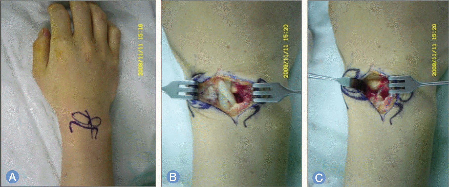
 | Fig. 1.
(A) Skin incition. (B) The extensor retinaculum between second and third extensor compartment is incised. (C) By placing retractors, the extensor carpi radialis longus tendon is retracted radially and the extensor carpi radialis brevis and the extensor pollicis longus tendons are retracted ulnarly. A longitudinal capsulotomy is performed along the long axis of the incision. |
 | Fig. 2.
(A) When a satisfactory reduction has been achieved, provisional fixation is obtained with two 0.045 K-wires which were inserted eccentrically from the dorsal ridge of the radius to slightly volar to the central axis of the scaphoid axis. (B, C) Using the K-wires as lever-arm, a scaphoid is volar flexed for the screw which is then inserted in the central axis. (D) Two K-wires which were fixed eccentrically is retracted volarly but not removed for additional stability. (E) In case of cyst formation, we can do additional bone graft through dorsal window after fixation of the scaphoid. |
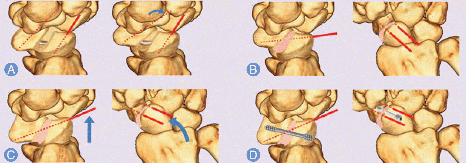
 | Fig. 3.
(A) 0.045 K-wire are inserted perpendicularly to the central axis of the scaphoid into the proximal and distal scaphoid fragments. Reduction of the nonunion site is attempted with assist of the joystick K-wires. (B) After insertion of the iliac bone graft into the gap, provisional fixation with two K-wires which were inserted eccentrically as the same procedure as simple nonunion. (C) Using the K-wires as a lever-arm, the scaphoid is volar flexed. (D) Herbert screw fixation is done in the central position with a free hand technique. |
Table 1.
Method of treatment


 PDF
PDF ePub
ePub Citation
 Print
Print


 XML Download