

 PDF
PDF ePub
ePub Citation
 Print
Print


Abstract
Intravenous pyogenic granuloma is a rare form of lobular capillary hemangioma and typically consists of an intraluminal polyp attached to the wall of a vein by a fibro-vascular stalk. It rarely occurs in the finger and its character is not enough to diagnosis clinically. Therefore, we report intravenous pyogenic granuloma which occurs in dorsal side of mid-phalanx with magnetic resonance imaging and pathological findings.
REFERENCES
1. Leyden JJ, Master GH. Oral cavity pyogenic granuloma. Arch Dermatol. 1973; 108:226–8.

2. Cooper PH, McAllister HA, Helwig EB. Intravenous pyogenic granuloma: a study of 18 cases. Am J Surg Pathol. 1979; 3:221–8.
3. Ghekiere O, Galant C, Vande Berg B. Intravenous pyogenic granuloma or intravenous lobular capillary hemangioma. Skeletal Radiol. 2005; 34:343–6.
4. Joethy J, Al Jajeh I, Tay SC. Intravenous pyogenic granuloma of the hand: a case report. Hand Surg. 2011; 16:87–9.
5. DiFazio F, Mogan J. Intravenous pyogenic granuloma of the hand. J Hand Surg Am. 1989; 14:310–2.
6. Qian LH, Hui YZ. Intravenous pyogenic granuloma: immunohistochemical consideration: a case report. Vasc Surg. 2001; 35:315–9.
7. Kamishima T, Hasegawa A, Kubota KC, et al. Intravenous pyogenic granuloma of the finger. Jpn J Radiol. 2009; 27:328–32.
8. Clearkin KP, Enzinger FM. Intravascular papillary endothelial hyperplasia. Arch Pathol Lab Med. 1976; 100:441–4.
9. Rosai J, Akerman LR. Intravenous atypical vascular proliferation: a cutaneous lesion simulating a malignant blood vessel tumor. Arch Dermatol. 1974; 109:714–7.
Fig. 1.
(A) Preoperative photogragh shows 1.5×0.5 cm palpable mass on the midphalanx level dorsal side of the fourth finger, left hand. (B) Intraoperative photogragh shows 1.6×0.6 cm sized well-lobulated pale mass like region in the vessel of mid phalanx dorsal side.
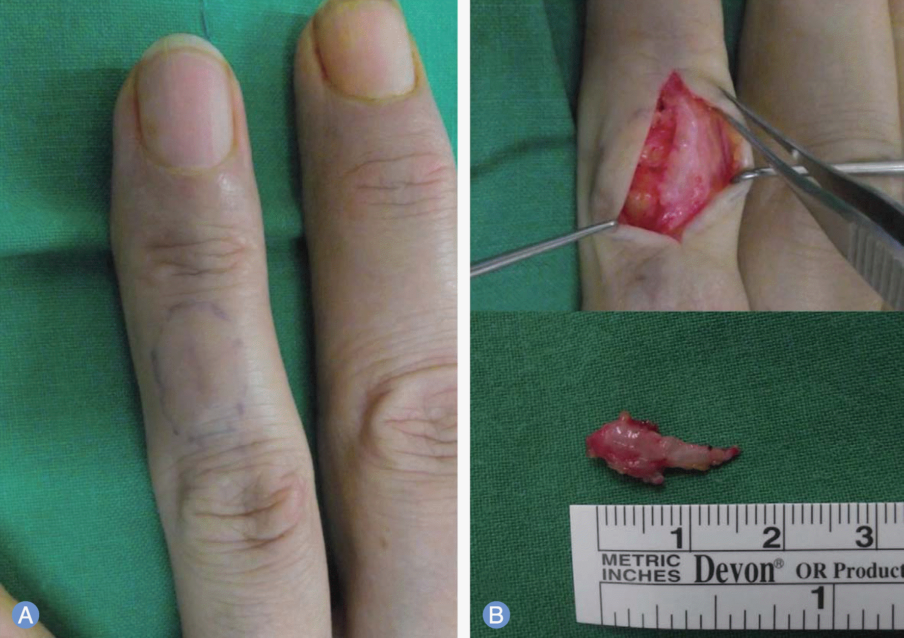
Fig. 2.
Magnetic resonance imaging. (A) Axial view T1-weighted image shows that the signal of the lesion is equivalent to that of adjacent skin. (B) Axial view T2-weighted image shows 1.2×0.4×0.7 cm-sized well-defined lobulating well-enhancing nodule at the subcutaneous layer of the middle phalanx level. (C) Axial and sagittal view T1-weighted image obtained after intravenous injection of gadolinium shows homogeneous enhancement of lesion.
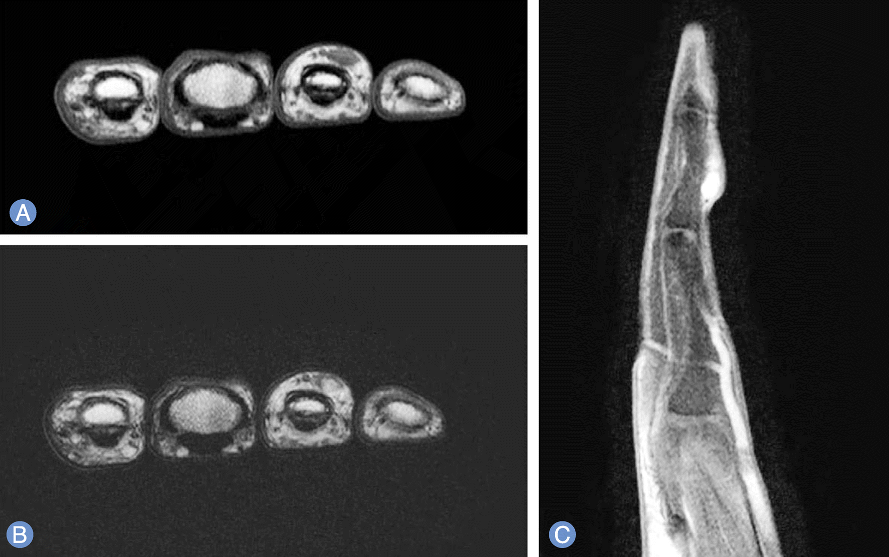
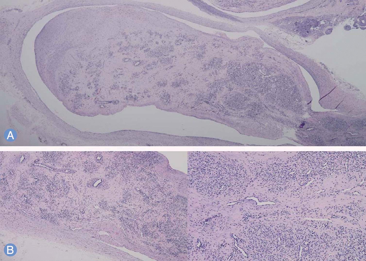
Fig. 3.
Histological photograph (H&E). (A) Subcutaneous nodule showing an intravenous polyp composed of lobules of capillaries separated by fibrous connective tissue attached to the wall of the vein by a fibrovascular stalk (×40). (B) Higher magnification shows monotonous endothelial hyperplasia and capillary proliferation (×200).
 XML Download