

 PDF
PDF ePub
ePub Citation
 Print
Print


Abstract
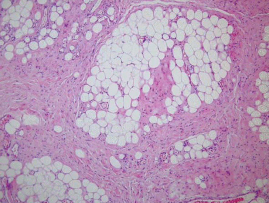
Lipofibromatous hamartoma is a very uncommon, benign tumor that involves diffuse infiltration of peripheral nerves by normal-appearing fibrous and adipose tissues. We repost a rare case of secondary carpal tunnel syndrome due to a lipofibromatous hamartoma of the median nerve with preoperative and post-operative magnetic resonance images.
Go to : 
REFERENCES
1. Mason ML. Proceedings of American Society for Surgery of the Hand: presentation of case. J Bone Joint Surg Am. 1953; 35:273–5.
2. Agarwal S, Haase SC. Lipofibromatous hamartoma of the median nerve. J Hand Surg Am. 2013; 38:392–7.

3. Tahiri Y, Xu L, Kanevsky J, Luc M. Lipofibromatous hamartoma of the median nerve: a comprehensive review and systematic approach to evaluation, diagnosis, and treatment. J Hand Surg Am. 2013; 38:2055–67.
4. Kim CH, Kim BK, Lim YH. Recurrent lipofibromatous hamartoma of the median nerve. J Korean Orthop Assoc. 2012; 47:316–20.
5. Park IH, Kim BK, Suh KJ, Jeon IH. Carpal tunnel syndrome due to lipofibromatous harmatoma of the median nerve: a case report. J Korean Soc Surg Hand. 2005; 10:79–82.
6. Al-Qattan MM. Lipofibromatous hamartoma of the median nerve and its associated conditions. J Hand Surg Br. 2001; 26:368–72.
7. Amadio PC, Reiman HM, Dobyns JH. Lipofibromatous hamartoma of nerve. J Hand Surg Am. 1988; 13:67–75.
8. Paletta FX, Senay LC Jr. Lipofibromatous hamartoma of median nerve and ulnar nerve: surgical treatment. Plast Reconstr Surg. 1981; 68:915–21.
9. Elsaidi GA, Wiesler ER. Lipofibromatous hamartoma of the median nerve: case presentation of MRI, ultrasound, electrodiagnostic, histologic, and surgical findings. Am J Orthop (Belle Mead NJ). 2004; 33:514–6.
10. Nardella D, Sohawon S, Carlier A. Lipofibromatous hamartoma of the median nerve: three case reports. J Plast Reconstr Aesthet Surg. 2009; 62:e314–7.
Go to :
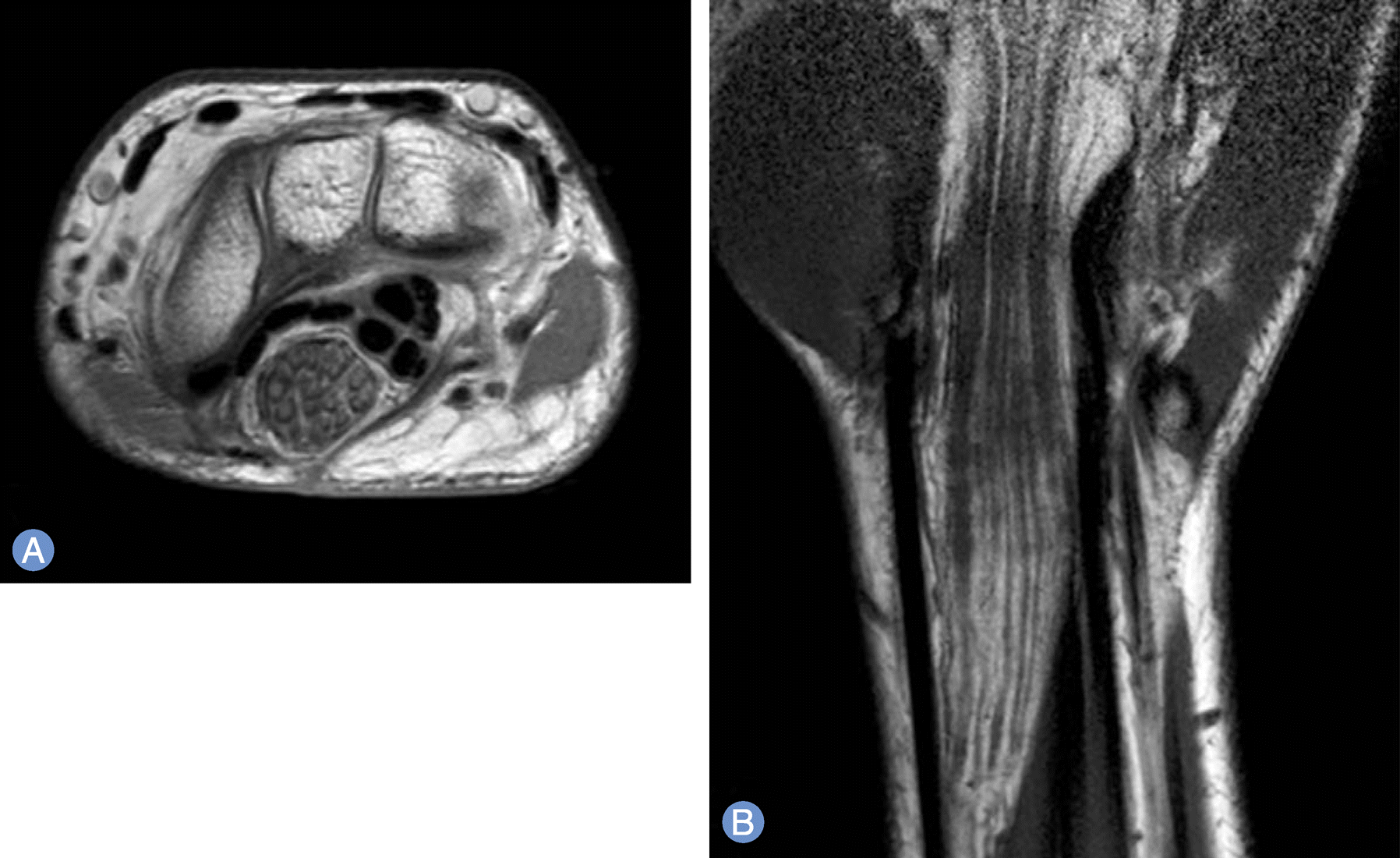
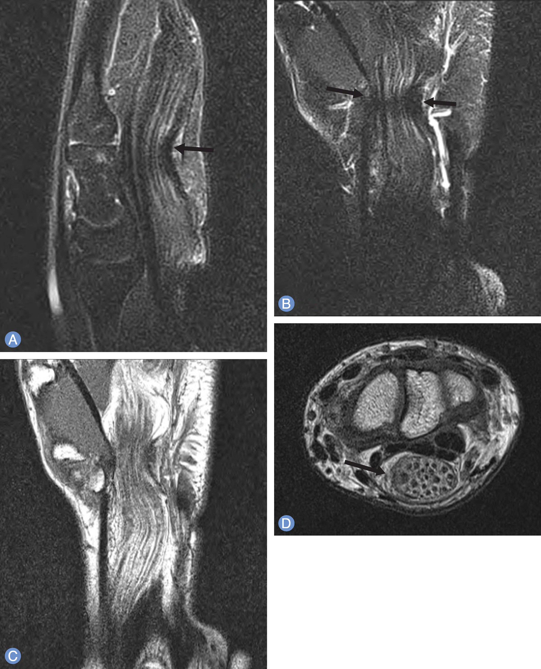 | Fig. 1.Magnetic resonance imaging findings of lipofibromatous hamartoma of the median nerve. (A) Sagittal- and (B) Coronal T2-weighted images showing the “hourglass like” appreance of the enlarged median nerve (arrows). (C) Coronal T1-weighted image showing the “spaghetti-like” appreance. (D) Axial T1 image showing the “coaxial cable like” appreance with displacementof flexor tendons (arrow). |
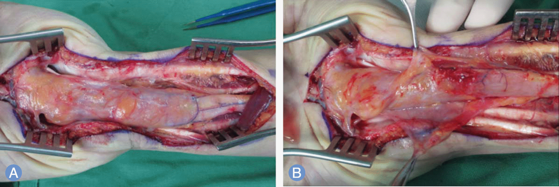 | Fig. 2.Intraoperative photographs. (A) The 12 × 2 × 2 cm fusiform median nerve with fibirofatty tissue is surrounded thick fibrous epineurium that is extended the distal forearm to palm. (B) Carpal tunnel release and epineurectomy were done. |
 XML Download