Abstract
An osteoid osteoma is a benign bone tumor. It is most commonly found in the femur and tibia but only 5% to 15% occurs in hand. Osteoid osteoma of carpal bone has vague nature of symptoms including spontaneous dull aching causing delayed diagnosis and the late treatment. We had a patient with an osteoid osteoma of the capitate bone presenting with tenosynovitis. We present clinical and radiological findings including magnetic resonance imaging, surgical result, and a review of the current literature.
Go to : 
REFERENCES
1. Marcuzzi A, Acciaro AL, Landi A. Osteoid osteoma of the hand and wrist. J Hand Surg Br. 2002; 27:440–3.

2. Bednar MS, Weiland AJ, Light TR. Osteoid osteoma of the upper extremity. Hand Clin. 1995; 11:211–21.

4. Murray PM, Berger RA, Inwards CY. Primary neoplasms of the carpal bones. J Hand Surg Am. 1999; 24:1008–13.

5. Edeiken J, DePalma AF, Hodes PJ. Osteoid osteoma. (Roentgenographic emphasis). Clin Orthop Relat Res. 1966; 49:201–6.

7. Campanacci M, Ruggieri P, Gasbarrini A, Ferraro A, Campanacci L. Osteoid osteoma. Direct visual identification and intralesional excision of the nidus with minimal removal of bone. J Bone Joint Surg Br. 1999; 81:814–20.
8. Rosenthal DI, Hornicek FJ, Wolfe MW, Jennings LC, Gebhardt MC, Mankin HJ. Percutaneous radiofrequency coagulation of osteoid osteoma compared with operative treatment. J Bone Joint Surg Am. 1998; 80:815–21.

Go to : 
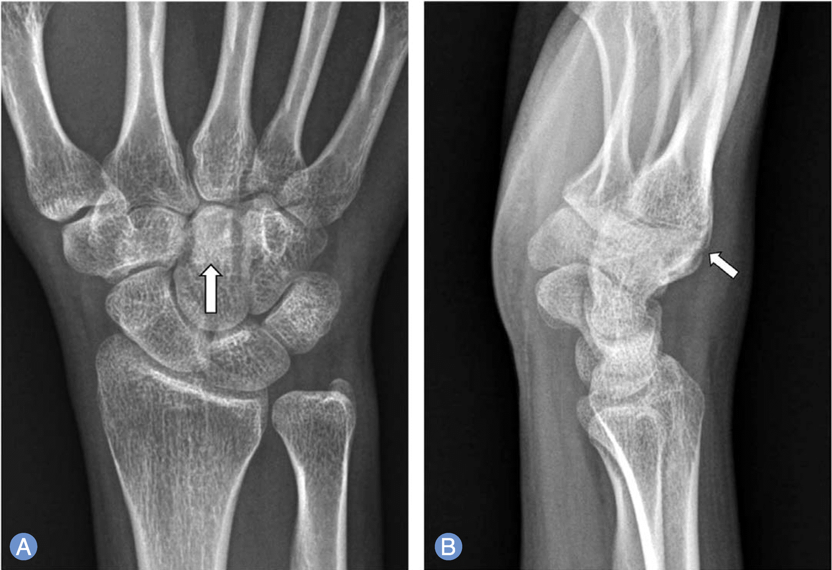
 | Fig. 2.
(A) Anteroposterior radiograph of the right wrist shows a sclerosis area (arrow) of the distal pole of the capitate. (B) On lateral radiograph, there is cortical disruption (arrow) at the dorsal capitate with the overlying soft tissue swelling. |
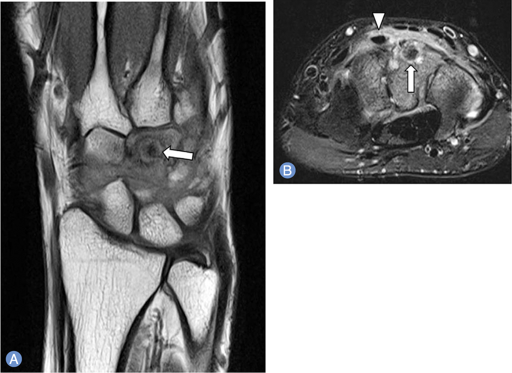
 | Fig. 3.
(A) Coronal T1-weighted magnetic resonance image (MRI) of right wrist shows a well circumscribed lesion (arrow) with central low signal in the capitate. MRI conditions were as follows: magnet strength 3.0T, fast spin echo, repetition time 670 msec, echo time 30 msec, field of view 11×11 cm, 2.0 thickness. (B) On axial T2-weighted with fat-suppression MR image obtained at the midcarpal level, this lesion (arrow) shows a hypointense center surrounded by hyperintense rim. There are extensive high signal suggesting bone marrow edema pattern in the remaining capitate, trapezoid and hamate with tenosynovitis of extensor carpi radialis brevis (arrow head). MRI conditions were as follows: magnet strength 3.0T, fast spin echo, repetition time 3,490 msec, echo time 68 msec, field of view 10 × 10 cm, 3.0 thickness. |


 PDF
PDF ePub
ePub Citation
 Print
Print


 XML Download