

 PDF
PDF ePub
ePub Citation
 Print
Print


Abstract
Purpose:
The purpose was to describe comparative analysis of the surgical outcome of percutaneous K-wire fixation of bony mallet fingers reduced with towel clip and 18-gauge needle.
Methods:
We analyzed the bony mallet finger patients with more than twelve months follow-up after percutaneous K-wire fixation. The patients were randomly divided into two groups. Eighteen fingers were treated with closed reduction using towel clip and 18 other fingers were treated with closed reduction using 18-gauge needle.
Results:
Radiographs showed bony union and no subluxation in all cases after K-wire removal. The average extension lag was 2.8°/1.9°, and range of motion of distal interphalangeal joint was 70.3°/75° respectively. According to Crawford's criteria, excellent results were obtained in 9/11 fingers, good results in 8/7 fingers, and poor result in 1/0 finger, respectively.
Go to : 
REFERENCES
1. Damron TA, Engber WD. Surgical treatment of mallet finger fractures by tension band technique. Clin Orthop Relat Res. 1994; (300):133–40.

2. Niechajev IA. Conservative and operative treatment of mallet finger. Plast Reconstr Surg. 1985; 76:580–5.
3. Bischoff R, Buechler U, De Roche R, Jupiter J. Clinical results of tension band fixation of avulsion fractures of the hand. J Hand Surg Am. 1994; 19:1019–26.
4. Choi JY, Jung HJ, Lee HJ, Son KM, Kim YH. Treatment of bony mallet finger: closed reduction using extension block K-wire. J Korean Fract Soc. 2004; 17:362–7.
5. Wehbe MA, Schneider LH. Mallet fractures. J Bone Joint Surg Am. 1984; 66:658–69.
6. Crawford GP. The molded polythene splint for mallet finger deformities. J Hand Surg Am. 1984; 9:231–7.
7. Hahn SH, Yang BK, Yi SR, Chung SW, Oh SJ. Clinical outcome of operative treatment for bony mallet finger. J Korean Soc Surg Hand. 2000; 5:68–74.
8. Hillman FE. New technique for treatment of mallet fingers and fractures of distal phalanx. J Am Med Assoc. 1956; 161:1135–8.
9. Kim SJ, Lee KH, Ha TS. Operative treatment of the mallet finger. J Korean Orthop Assoc. 1992; 27:796–801.
10. Pratt DR, Bunnell S, Howard LD Jr. Mallet finger; classification and methods of treatment. Am J Surg. 1957; 93:573–8.
11. Okafor B, Mbubaegbu C, Munshi I, Williams DJ. Mallet deformity of the finger. Five-year follow-up of conservative treatment. J Bone Joint Surg Br. 1997; 79:544–7.
12. Stern PJ, Kastrup JJ. Complications and prognosis of treatment of mallet finger. J Hand Surg Am. 1988; 13:329–34.
13. Lubahn JD. Mallet finger fractures: a comparison of open and closed technique. J Hand Surg Am. 1989; 14:394–6.
14. Stark HH. Troublesome fractures and dislocations of the hand. American Academy of Orthopedic Surgeons , editor. Instructional course lectures. Vol 19. St. Louis: Mosby;1970. p. 130–49.
15. Takami H, Takahashi S, Ando M. Operative treatment of mallet finger due to intra-articular fracture of the distal phalanx. Arch Orthop Trauma Surg. 2000; 120:9–13.
16. King HJ, Shin SJ, Kang ES. Complications of operative treatment for mallet fractures of the distal phalanx. J Hand Surg Br. 2001; 26:28–31.
17. Ishiguro T, Inoue K, Matsubayashi N, Ito Y, Hashizume N. A new method of closed reduction for mallet fractures. Cent Jpn Orthop Traumatol. 1988; 31:2049–51.
18. Han CS, Chung DW, Jeong BO, et al. Percutaneous Kirschner wire fixation of acute mallet fractures percutaneousely reduced by towel clip. J Korean Fract Soc. 2009; 22:283–7.
19. Han KJ, Moon JH, Chung NS, Kang SY. Treatment of mallet fracture of the fingers by percutaneous reduction and pinning. J Korean Soc Surg Hand. 2003; 8:153–7.
Go to :
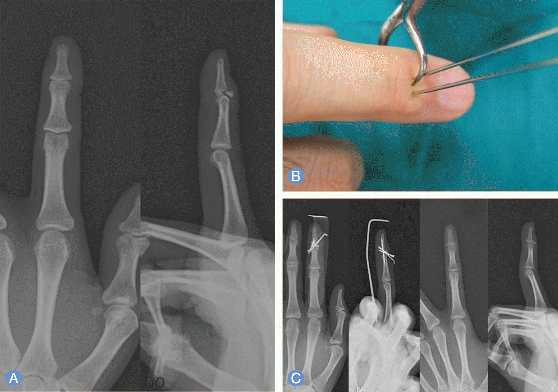 | Fig. 1.A 24-year-old man has a bony mallet finger injury of index finger. (A) The initial anterioposterior and lateral X-ray show displaced dorsal fragment and articular involvement more than 30% without subluxation. (B) Percutaneous pinning with towel clip reduction is performed. (C) At postopertive 12 months, the fracture is united and the patient has a range of motion from 0 to 75 degree. |
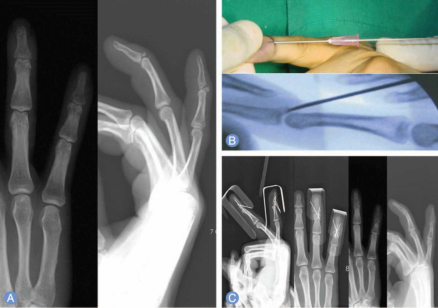 | Fig. 2.A 36-year-old man has a bony mallet finger injury of the 4, 5th finger. (A) The initial anteriorposterior and lateral X-ray show a displaced dorsal fragment and articular involvement more than 30% without subluxation. (B) Percutaneous pinning with towel clip reduction is performed. (C) At postoperative 12 months, the fracture is united and the patient has a range of motion from 5 to 78 degree on the 3th finger and 0 to 78 degree on the 4th finger. |
Table 1.
Summary of group A cases
Table 2.
Summary of group B cases
Table 3.
Whebe and Schneider's classification
 XML Download