Journal List > J Korean Assoc Oral Maxillofac Surg > v.44(5) > 1106350

Farzin, Taghva, and Babooie: Comparison of temporomandibular disorders between menopausal and non-menopausal women

Abstract

Objectives

Hormonal changes during menopause alter a woman's susceptibility to some disorders. Information regarding the prevalence of temporomandibular disorder (TMD) in menopausal women is limited in the literature. In this study, the prevalence and severity of TMDs were compared between menopausal and non-menopausal women.

Materials and Methods

The study included 140 women (69 premenopausal and 71 postmenopausal) 45 to 55 years of age that were examined in Shiraz Dental School, Shiraz in Iran. The Helkimo clinical dysfunction index (Di) was used to evaluate temporomandibular joint (TMJ) dysfunction. The data were analyzed using chi-square and Fisher's exact tests.

Results

Occurrence of TMD was significantly higher in menopausal than non-menopausal women (P<0.001). All the TMD criteria based on Helkimo Di except range of mandibular movement were significantly more common in menopausal women. The range of mandibular movement was not significantly different between menopausal and non-menopausal women (P=0.178).

Conclusion

The results from this study show that TMD can be considered more common and severe in menopausal than non-menopausal women. This finding indicates that, similar to other conditions in menopausal women such as arthritis and osteoporosis, TMD should be taken into consideration by dental and medical professionals.

I. Introduction

Temporomandibular disorders (TMDs) refer to a group of musculoskeletal disorders affecting the masticatory muscle and/or the temporomandibular joint (TMJ)1. Reportedly, 33% to 86% of individuals exhibit at least one sign of TMD, and 16% to 59% have at least one symptom234. Although the etiology of TMDs is not completely known, different factors such as occlusion, parafunction, emotional stress, hormones, physical trauma, microtrauma to the teeth, joint hypermobility, and dental treatments requiring extensive chair time have been considered in the etiology of TMDs in various studies5678910. Estrogen and progesterone receptors were detected in the human TMJ disc, suggesting that changes in reproductive hormones may play a role in the pathogenesis of joint disorders11. Therefore, hormonal changes in women during menopause may alter their susceptibility to joint disease.
Information regarding the role of female sexual hormones in the etiology of joint diseases is contradictory in the literature. Some studies have shown that estrogen is necessary for an intact TMJ structure121314151617. Madani et al.12 found the level of progesterone to be lower in a group of women with TMJ clicking; however, the estrogen level was the same in the patient and control groups. In another study, Abubaker et al.13 found that estrogen is necessary for development of collagen and protein in the structure of TMJ in rats. However, in other studies, a higher prevalence of TMDs with increased estrogen level was reported1617.
Several indices have been developed to assess the prevalence and severity of signs and symptoms of TMDs181920. One of the most feasible was developed by Helkimo20; the Helkimo clinical dysfunction index (Di) assesses the prevalence and severity of signs and symptoms in patients with TMD20.
A significant reduction in gonadal hormones occurs in postmenopausal women and is responsible for many disorders such as cardiovascular disease and neurologic disease2122. Although the relationships between menopause and certain joint diseases such as postmenopausal osteoarthritis and rheumatoid arthritis are known, information regarding the prevalence of TMDs in menopausal women is limited in the litrature2324. Therefore, in the present study, the prevalence and severity of TMDs were assessed using the Helkimo Di in menopausal and non-menopausal women who were referred to Shiraz Dental School for a consultation from February to November 2015.

II. Materials and Methods

This study was approved by the Ethical Committee of Shiraz University of Medical Sciences (approval no. 1479). The informed consent was waived for this retrospective study. A cross-sectional study was performed in 140 women 45 to 55 years of age who were referred to the prosthodontics department of Shiraz Dental School (Shiraz, Iran) from February to November 2013. All patients were informed regarding the study and agreed to participate. Menopausal women whose latest menstruation occurred at least 1 year prior or had not used medication containing sexual hormones for the last 3 months were selected. Among non-menopausal women, females who had a normal menstrual cycle and did not use drugs containing sexual hormones for the last 3 months were selected. All the subjects were generally healthy and did not have history of trauma or surgery in the TMJ region.
All participants were evaluated using the Helkimo clinical Di, which has five subscales for assessing clinical signs of TMDs (mobility, TMJ function, muscle pain, TMJ pain, and pain during mandibular movement index).(Table 1)
Finally, the values of the five indices were summed, and patients were classified into 4 groups according to clinical Di.(Table 1)
Clinical examination was performed twice by a trained dental student. All patients were examined on a dental unit using the same calipers and under the same light. Results of the clinical examination were recorded on a standardized chart according to Helkimo20. The data were analyzed using PASW Statistics (ver. 18.0; IBM Co., Armonk, NY, USA) and chi-square and Fisher's exact tests.

III. Results

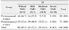
A total of 140 women, 69 premenopausal (49.3%) and 71 postmenopausal (50.7%), were recruited for this cross-sectional study. The mean age of the menopausal women was 46.87 years, and that of non-menopausal women was 51.34 years.
Based on the Helkimo Di, 49.3% of subjects had no signs of TMD (Di 0), and 50.7% suffered from some degree of TMD (Di I, II, or III). Among menopausal women, 40.8% had mild TMD (Di I), 21.1% moderate (Di II), and 5.6% showed signs and symptoms of severe TMD (Di III). Among non-menopausal women, 23.2% had mild TMD (Di I), 7.2% moderate (Di II), and 2.9% suffered from severe TMD (Di III).(Table 2) TMD occurrence was significantly higher in menopausal women than non-menopausal women (P<0.001). The odds ratio of TMD occurrence between menopausal and non-menopausal women was 4.17.
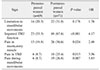
The prevalence of clinical signs and symptoms of TMDs in menopausal women is summarized in Table 3. Among these clinical signs and symptoms, pain in the TMJ and pain during mandibular movements were the least prevalent findings in menopausal and non-menopausal women, respectively. The most common finding in both groups was minor problems in TMJ function and was approximately 2 times more common in menopausal women than non-menopausal women. All TMD criteria based on Helkimo Di, except range of mandibular movement, were significantly more common in menopausal women.

IV. Discussion

Based on the results from this study, occurrence of TMDs was significantly more frequent in menopausal women than non-menopausal women; except for minor problems in mandibular movement (P=0.178), all other Helkimo indices were significantly higher in menopausal women. LeResche et al.25 reported the highest TMD pain level in women when estrogen was at its lowest level during the menstrual cycle. In another study, Haskin et al.26 showed an inverse relationship between blood estrogen level and joint pain, which is in agreement with the current study results.
Some studies272829 have shown that estrogen has an important role in the development of TMJ structure. Yasuoka et al.15 showed that ovarian hormone deficiency in ovariectomized rats resulted in destructive changes to the TMJ structure, which could be prevented by estradiol replacement. Reportedly, estrogen deficiency in rats during puberty changes serum calcitonin and parathyroid hormone levels, causing alterations in rat TMJ structure14. In another study, Abubaker et al.13 showed the necessity of estrogen for development of collagen and protein in the structure of the TMJ disc of the rat. Regarding these findings, reduction in estrogen level during menopause may result in less development of the TMJ structure; therefore, increased severity and prevalence of TMDs in menopausal women could be expected based on the results from these studies.
In addition, anti-nociceptive and inflammatory effects of estradiol, the most common form of estrogen, have been demonstrated in several studies2728. Fischer et al.27 showed that nociceptive behavior induced by injection of formalin or glutamate into the TMJ is higher at low serum estradiol level in female rats, and vice versa. Torres-Chávez et al.28 found that estradiol reduces plasma extravasations and neutrophil migration, two parameters of TMJ inflammation. This anti-inflammatory effect of estradiol reduces damage to the TMJ.
Results of several studies have shown that reduction in plasma estrogen level reduced the inflammatory factors in TMJ293031. For example, Yun et al.29 found that estrogen could stimulate expression of proinflammatory cytokines such as interleukin (IL)-6, IL-8, and IL-18, which would increase TMD. Contrary to the present study results, several studies reported TMD increase when sexual hormone levels increase1632. Landi et al.16 found that patients with TMD had significantly higher serum level of estrogen. LeResche et al.32 showed that TMD was less frequent in menopausal women, and use of postmenopausal estrogen increased TMD in women. However, a later study did not show any relationship between estrogen replacement therapy and increased risk of TMD in women33. Dose-dependent effect of estrogen in the physiopathology of TMDs might explain this contrast. In other words, a physiological level of estrogen is necessary for proper development of the TMJ structure, but higher levels could result in inflammation and TMD34. Another reason might be the different criteria used in these studies and current investigation for diagnosis of TMDs1131.
One limitation of this study was lack of measurement of exact hormonal levels in the study participants. Another limitation was the relatively small study group. Although, the number of subjects was adequate for statistical analysis, future studies with a larger cohort of women are recommended for more feasible results.

V. Conclusion

Within the limitations of this study, the prevalence and severity of TMDs were higher in menopausal women than non-menopausal women. Therefore, TMDs, like other common conditions such as arthritis and osteoporosis that affect menopausal women, should be taken into consideration by dental and medical professionals.

Figures and Tables

Table 1

Clinical dysfunction index

jkaoms-44-232-i001

(TMJ: temporomandibular joint, MMO: maximal mouth opening, Di: dysfunction index, TMD: temporomandibular disorder)

1>40 mm=0 point; 30–39 mm=1 point; <30 mm=5 points; 2≥7 mm=0 point; 4–6 mm=1 point; 0–3 mm=5 points.

Total dysfunction score (sum A–E): 0=Di 0 (absence of clinical symptoms); 1–4=Di I (minor TMD); 5–9=Di II (moderate TMD); 10–25=Di III (severe TMD).

Table 2

Comparison of degree of dysfunction index (Di) in postmenopausal and premenopausal women

jkaoms-44-232-i002

(TMD: temporomandibular disorder)

Values are presented as number (%).

Table 3

Prevalence of signs and symptoms of TMD dysfunction in 140 menopausal and non-menopausal women based on Helkimo index

jkaoms-44-232-i003

(TMD: temporomandibular disorder, OR: odds ratio, TMJ: temporomandibular joint)

Acknowledgements

This manuscript is based on the thesis by Dr. Moslem Babooie (no. 1479).
The authors thank the Vice-Chancellor of Shiraz University of Medical Sciences for supporting this research. The authors also thank Dr. Vosoughi of the Dental Research Development Center from the School of Dentistry for the statistical analysis and Dr. Shahram Hamedani (DDS, MSc) for his suggestions and editorial assistance in the manuscript.

Notes

Authors' Contributions M.F. participated in the study design. M.T. wrote the manuscript. M.B. participated in data collection.

Ethics Approval and Consent to Participate This study was approved by the Ethical Committee of Shiraz University of Medical Sciences (approval no. 1479). The informed consent was waived for this retrospective study.

Conflict of Interest No potential conflict of interest relevant to this article was reported.

References

1. Okeson JP. Management of temporomandibular disorders and occlusion. St. Louis: Mosby Elsevier;2013. p. 129.
2. Ebrahimi M, Dashti H, Mehrabkhani M, Arghavani M, Daneshvar-Mozafari A. Temporomandibular disorders and related factors in a group of Iranian adolescents: a cross-sectional survey. J Dent Res Dent Clin Dent Prospects. 2011; 5:123–127.
3. De Leeuw R. [Orofacial pain: guidelines for assessment, diagnosis, and management]. 4th ed. São Paulo: Quintessence;2010. Portuguese.
4. Carlsson G, LeResche L. Epidemiology of temporomandibular disorders. In : Sessle B, Bryant P, Dionne R, editors. Progress in pain research and management. Seattle: International Association for the Study of Pain;1995. p. 211–226.
5. Poveda-Roda R, Bagán JV, Díaz-Fernández JM, Hernández-Bazán S, Jimenez-Soriano Y. Review of temporomandibular joint pathology. Part I: classification, epidemiology and risk factors. Med Oral Patol Oral Cir Bucal. 2007; 12:E292–E298.
6. Sari S, Sonmez H. Investigation of the relationship between oral parafunctions and temporomandibular joint dysfunction in Turkish children with mixed and permanent dentition. J Oral Rehabil. 2002; 29:108–112.
crossref
7. Winocur E, Littner D, Adams I, Gavish A. Oral habits and their association with signs and symptoms of temporomandibular disorders in adolescents: a gender comparison. Oral Surg Oral Med Oral Pathol Oral Radiol Endod. 2006; 102:482–487.
crossref
8. Emodi-Perlman A, Eli I, Friedman-Rubin P, Goldsmith C, Reiter S, Winocur E. Bruxism, oral parafunctions, anamnestic and clinical findings of temporomandibular disorders in children. J Oral Rehabil. 2012; 39:126–135.
crossref
9. Clark GT. Etiologic theory and the prevention of temporomandibular disorders. Adv Dent Res. 1991; 5:60–66.
crossref
10. Huang GJ, LeResche L, Critchlow CW, Martin MD, Drangsholt MT. Risk factors for diagnostic subgroups of painful temporomandibular disorders (TMD). J Dent Res. 2002; 81:284–288.
crossref
11. Abubaker AO, Raslan WF, Sotereanos G. Estrogen and progester-one receptors in temporomandibular joint disc of symptomatic and asymptomatic persons: a preliminary study. J Oral Maxillofac Surg. 1993; 51:1096–1100.
12. Madani AS, Shamsian AA, Hedayati-Moghaddam MR, Fathi-Moghadam F, Sabooni MR, Mirmortazavi A, et al. A cross-sectional study of the relationship between serum sexual hormone levels and internal derangement of temporomandibular joint. J Oral Rehabil. 2013; 40:569–573.
crossref
13. Abubaker AO, Hebda PC, Gunsolley JN. Effects of sex hormones on protein and collagen content of the temporomandibular joint disc of the rat. J Oral Maxillofac Surg. 1996; 54:721–727.
crossref
14. Okuda T, Yasuoka T, Nakashima M, Oka N. The effect of ovariectomy on the temporomandibular joints of growing rats. J Oral Maxillofac Surg. 1996; 54:1201–1210.
crossref
15. Yasuoka T, Nakashima M, Okuda T, Tatematsu N. Effect of estrogen replacement on temporomandibular joint remodeling in ovariectomized rats. J Oral Maxillofac Surg. 2000; 58:189–196.
crossref
16. Landi N, Manfredini D, Lombardi I, Casarosa E, Boseo M. 17-beta estradiol and progesterone serum levels in temporomandibular disorder patients. Minerva Stomatol. 2004; 53:651–660.
17. Smith YR, Stohler CS, Nichols TE, Bueller JA, Koeppe RA, Zubieta JK. Pronociceptive and antinociceptive effects of estradiol through endogenous opioid neurotransmission in women. J Neurosci. 2006; 26:5777–5785.
crossref
18. Dworkin SF, LeResche L. Research diagnostic criteria for temporomandibular disorders: review, criteria, examinations and specifications, critique. J Craniomandib Disord. 1992; 6:301–355.
19. Fricton JR, Schiffman EL. Reliability of a craniomandibular. J Dent Res. 1986; 65:1359–1364.
20. Helkimo M. Studies on function and dysfunction of the masticatory system. IV. Age and sex distribution of symptoms of dysfunction of the masticatory system in Lapps in the north of Finland. Acta Odontol Scand. 1974; 32:255–267.
21. Dosi R, Bhatt N, Shah P, Patell R. Cardiovascular disease and menopause. J Clin Diagn Res. 2014; 8:62–64.
22. Jamshed N, Ozair FF, Aggarwal P, Ekka M. Alzheimer disease in post-menopausal women: intervene in the critical window period. J Midlife Health. 2014; 5:38–40.
crossref
23. Bay-Jensen AC, Slagboom E, Chen-An P, Alexandersen P, Qvist P, Christiansen C, et al. Role of hormones in cartilage and joint metabolism: understanding an unhealthy metabolic phenotype in osteoarthritis. Menopause. 2013; 20:578–586.
24. Macovei L, Ancuţa C, Belibou C, Chirieac R. [Bone mineral density in patients with rheumatoid arthritis]. Rev Med Chir Soc Med Nat Iasi. 2011; 115:723–730. Romanian.
25. LeResche L, Mancl L, Sherman JJ, Gandara B, Dworkin SF. Changes in temporomandibular pain and other symptoms across the menstrual cycle. Pain. 2003; 106:253–261.
crossref
26. Haskin CL, Milam SB, Cameron IL. Pathogenesis of degenerative joint disease in the human temporomandibular joint. Crit Rev Oral Biol Med. 1995; 6:248–277.
crossref
27. Fischer L, Torres-Chavez KE, Clemente-Napimoga JT, Jorge D, Arsati F, de Arruda, et al. The influence of sex and ovarian hormones on temporomandibular joint nociception in rats. J Pain. 2008; 9:630–638.
crossref
28. Torres-Chávez KE, Sanfins JM, Clemente-Napimoga JT, Pelegrini-Da-Silva A, Parada CA, Fischer L, et al. Effect of gonadal steroid hormones on formalin-induced temporomandibular joint inflammation. Eur J Pain. 2012; 16:204–216.
crossref
29. Yun KI, Chae CH, Lee CW. Effect of estrogen on the expression of cytokines of the temporomandibular joint cartilage cells of the mouse. J Oral Maxillofac Surg. 2008; 66:882–887.
crossref
30. Flake NM, Hermanstyne TO, Gold MS. Testosterone and estrogen have opposing actions on inflammation-induced plasma extravasation in the rat temporomandibular joint. Am J Physiol Regul Integr Comp Physiol. 2006; 291:R343–R348.
crossref
31. Craft RM. Modulation of pain by estrogens. Pain. 2007; 132:Suppl 1. S3–S12.
crossref
32. LeResche L, Saunders K, Von Korff MR, Barlow W, Dworkin SF. Use of exogenous hormones and risk of temporomandibular disorder pain. Pain. 1997; 69:153–160.
crossref
33. Nekora-Azak A, Evlioglu G, Ceyhan A, Keskin H, Berkman S, Issever H. Estrogen replacement therapy among postmenopausal women and its effects on signs and symptoms of temporomandibular disorders. Cranio. 2008; 26:211–215.
crossref
34. Wu YW, Kou XX, Bi RY, Xu W, Wang KW, Gan YH, et al. Hippocampal nerve growth factor potentiated by 17β-estradiol and involved in allodynia of inflamed TMJ in rat. J Pain. 2012; 13:555–563.
crossref
TOOLS
ORCID iDs

Mitra Farzin
https://orcid.org/0000-0003-2352-7211

Masumeh Taghva
https://orcid.org/0000-0001-6662-2257

Moslem Babooie
https://orcid.org/0000-0002-6206-9973

Similar articles