

 PDF
PDF ePub
ePub Citation
 Print
Print


I. Introduction
Bisphosphonate-related osteonecrosis of the jaw (BRONJ) was renamed to medication-related osteonecrosis of the jaw (MRONJ) as the incidence of jaw necrosis is increasing in relation to other bone resorption inhibitors or angiogenesis inhibitors1. Affiliated drugs are currently being used in various clinical applications. Oral administration of bisphosphonates is mainly performed for osteoporosis and osteogenesis2. Intravenous bisphosphonates are used for hypercalcemia associated with malignant disease and management of skeletal complications and osteolytic lesions in osteopathic cancer patients and osteoporosis patients345. Another bone resorption inhibitor, denosumab, is administered orally or subcutaneously to inhibit skeletal complications in bone metastatic lesion and to reduce vertebral and hip fractures in osteoporosis patients67. Pathophysiologically, the exact mechanism of MRONJ development remains unclear, but it has been reported to potentially be due to excessive inhibition of jaw metabolic processes, infection and inflammation, inhibition of angiogenesis, soft tissue toxicity, immune system abnormalities, and accumulation of microfractures8910111213.
As the number of MRONJ patients continues to increase, related research is actively being conducted. The American Association of Oral and Maxillofacial Surgeons (AAOMS) in 2014 and the Korean Society for Bone and Mineral Research (KSBMR) and the Korean Association of Oral and Maxillofacial Surgeons (KAOMS) in 2015 published position papers on MRONJ114. According to these studies, MRONJ can be diagnosed if (1) there is a history of using a bone resorption inhibitor or an angiogenesis inhibitor and if (2) there is no history of radiation therapy to the jaw, exposure of the jaw, or oral or extraoral fistula lasting more than eight weeks. In addition, MRONJ can be divided into stages according to progression. In stage 0, there are no clinical symptoms of osteonecrosis but a nonspecific symptom. In stage 1, necrotic bone is exposed, but there is no evidence of symptoms or infection. In stage 2, there is osteonecrosis with symptoms and infection. Lastly, stage 3 involves the same stage 2 findings with necrotic bone beyond the alveolar bone (i.e., mandibular inferior border or maxillary sinus), pathological fractures, or extraoral fistula (i.e., orocutaneous fistula or oronasal and oroantral fistula).
The risk factors for MRONJ can be divided into systemic factors and local factors. Systemic factors include the duration of related-medication use, use of steroids, age, diabetes, and genetic factors, while local factors include invasive oral surgery, thin mucosa, and periodontal disease151617. Depending on the symptoms and progression of the disease, conservative treatment including pain control, antibiotics, antibacterial gaggle, and various surgical treatments for removing necrotic tissue can be performed. Surgical treatment has been reported to yield a higher success rate, although failure may lead to advanced necrosis1819.
Studies on factors affecting recurrence of MRONJ are rare in the South Korean population. Several investigations have suggested duration of administration of related medications, presence of bacterial infections in necrotic areas, and methods of treatment used as factors in recurrence2021. Therefore, pathophysiologic causes and risk factors of MRONJ as well as factors affecting recurrence should be examined. The purpose of the present study was to investigate the demographic and clinical characteristics of patients with MRONJ and to elucidate factors affecting recurrence following surgical treatment.
II. Materials and Methods
This study was approved by the Institutional Review Board of Korea University Anam Hospital (Seoul, Korea). From 2013 to 2017, a total of 51 patients who were diagnosed with MRONJ in the Department of Oral and Maxillofacial Surgery, Korea University Anam Hospital were enrolled in the present study. The diagnosis of MRONJ was based on the AAOMS 2014 position paper1. Demographic, clinical, and treatment outcomes data were analyzed by retrospective chart review. When referring to local hospitals, referrals were also consulted. In the demographic analysis, the sex and age of the patients were examined. In the clinical characteristics analysis, the type of MRONJ-inducing drug used and the method and duration of administration were examined. We also investigated the MRONJ-inducing dental treatment performed as well as the site, stage, treatment course, and results of MRONJ and furthermore examined the size of the lesion by way of panoramic radiographic study. The closure method was divided into two categories: primary closure and secondary healing. Primary closure was performed without tension by incision on the periosteum of the mucoperiosteal flap after removal of the lesion. Secondary healing was performed with antibiotic gauze packing (Furacin gauze) with subsequent gauze and dressing changes performed every two to three days following removal of the lesion. Recurrence of MRONJ was defined as multiple infections and severe pain not responding to surgical treatment. Statistical analysis using the IBM SPSS Statistics (ver. 22; IBM Co., Armonk, NY, USA) was performed to analyze the factors affecting recurrence in patients who received surgical treatment. Fisher's exact test and the chi-square test were also employed for statistical analysis.
III. Results
1. Demographic analysis
Of the total 51 patients, three patients (5.9%) were male and 48 patients (94.1%) were female, and the mean patient age was 76.1±9.5 years (range, 45–92 years). The prevalence rate of MRONJ increased with age, with four patients (7.8%) being younger than 60 years, eight patients (15.7%) being between 61 years and 70 years, 21 patients (41.2%) being between 71 years and 80 years, and 18 patients (35.3%) being older than 80 years.
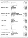
2. Clinical characteristics (Table 1)
1) MRONJ-inducing medications
Of the 51 patients, six patients were taking osteoporosis medication, but the type and duration of the drug were not specified and were excluded. Among the 45 patients, alendronate was the most common (25 patients, 55.6%) medication noted, followed by ibandronate (9 patients, 20.0%), risedronate (7 patients, 15.6%), zoledronate (3 patients, 6.7%), and pamidronate (1 patient, 2.2%). Forty patients (88.9%) had been given their medications orally, while five patients (11.1%) had undergone intravenous administration. The mean duration of drug use was 61.1±42.9 months (range, 6–240 months). A total of 10 patients had experienced a drug holiday prior to the dental treatment that induced MRONJ, with a mean length of 6.8±7.0 months.
2) Medical history associated with MRONJ
Of the total 51 patients, including those with multiple diseases, 37 patients (72.5%) had osteoporosis and 14 patients (27.5%) had diabetes mellitus. Additionally, five patients (9.8%) had been treated with steroids due to lupus, rheumatism, or adrenal insufficiency, and six patients (11.8%) had cancer. Patients without associated medical history were taking prophylactic osteoporosis medications.
3) Disease site, cause, stage, and size
Two different disease sites in a single patient were counted as two cases. With regard to site, of the total 56 cases, mandibular lesions were 2.7 times more prevalent, found in 41 cases (73.2%) versus the maxilla in 15 cases (26.8%). Forty-eight cases (85.7%) of posterior teeth (premolar-molar) made this presentation six times more prevalent than the eight cases (14.3%) of anterior teeth (incisor-canine). Regarding cause, extraction (39 cases, 69.6%) was the most common cause, with ill-fitting denture (8 cases, 14.3%); implant installation (6 cases, 10.7%); and other causes such as periodontal disease, alveoloplasty, and/or trauma (3 cases, 5.4%) also being noted. With respect to disease stage, stage 2 (31 cases, 55.4%) was the most common, while there were six cases (10.7%) of stage 0, 15 cases (26.8%) of stage 1, and four cases (7.1%) of stage 3. For lesion size, the width and height of both bone destruction and sclerosis were analyzed on panoramic images. In 41 cases showing bone destruction, the mean size was 20.0±6.5 mm in width and 12.6±5.2 mm in height.
4) Treatment outcome
Conservative treatment was performed in eight patients (15.7%), and surgical treatment was performed in 43 patients (84.3%). A total of 54 surgical treatments were completed, including in those who had relapsed and required surgery again. Of these, general anesthesia was used in 24 cases (44.4%), and local anesthesia was used in 30 cases (55.6%). When classified according to the closure method, 34 cases (63.0%) involved primary suture, and 20 cases (37.0%) involved secondary healing. Recurrence occurred in 13 cases, a rate of 24.1%. The mean follow-up period was 9.6±10.8 months.
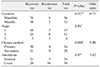
3. Recurrence factors
In the study, we analyzed whether there was a difference in recurrence rate according to site, stage, closure, and anesthesia method. Of the 48 MRONJ cases in 43 surgically treated patients, including two cases of multisite disease in a single patient, 8 of the 36 mandible cases (22.2%) and 2 of 12 maxilla cases (16.7%) involved relapse. There was no statistical difference in recurrence rate according to site. Additionally, 3 of 13 stage 1 cases (23.1%), 6 of 31 stage 2 cases (19.4%), and 1 of 4 stage 3 cases (25.0%) demonstrated relapse, with no statistical difference in recurrence rate according to stage.
Among 54 cases of surgical treatment, 4 of 34 primary closure cases (11.8%) and 9 of 20 secondary healing cases (45.0%) experienced relapse, and there was a statistically significant difference in recurrence rate according to closure method (P=0.008). Of the 24 cases, 3 general anesthesia cases (12.5%) and 10 of 30 local anesthesia cases (33.3%) demonstrated relapse. There was a difference in recurrence rate according to anesthesia, although no statistical significance was found.(Table 2)
IV. Discussion
Various systemic and local factors are known to be risk indicators for MRONJ. Previously, the occurrence has demonstrated an increase in patients older than 65 years of age22, and similar results were found in the present work. The prevalence of MRONJ in the patients who received drugs orally was 0.00104% to 0.00169%232425, whereas higher incidence of MRONJ was noted on the patients with intravenous zoledronate (0.017%) and denosumab (0.04%), respectively2627. It has been reported that the risk of MRONJ is increased specifically in diabetic patients, which is due to decreases in bone quality associated with capillary ischemia, vascular endothelial function, osteoblast and bone cell death, immune cell function, and increase in inflammation16. The use of steroids may also be a risk factor for MRONJ development due to decreased immune cells, delayed wound healing associated with steroid use, and worsening oral inflammation28. In an animal study, concurrent use of bisphosphonates and steroids increased the incidence of BRONJ29. MRONJ has additionally been reported in patients with cancer such as breast cancer and multiple myeloma3031. As a local risk factor, alveolar bone surgery is considered to be the main cause of MRONJ. In the present study, extraction was the most common cause of MRONJ (70.6%), as in previous studies (70.6%)283233. Other invasive alveolar bone surgeries such as implant installation, endodontic treatment, and periodontal surgery have not been as adequately investigated as a cause of MRONJ, but they are considered to be similar risk factors for extraction. In this study, MRONJ was 2.7 times more common in the mandible than in the maxilla in 73.2% of patients, and the posterior part was more common than the anterior part because the lesion might constitute a more complex vascular network and a more abundant blood supply in the maxilla than in the mandible. Notably, previous studies have reported similar results28.
In a previous study that analyzed panoramic radiographs of patients with MRONJ, it was reported that osseous sclerosis, lamina dura thickening, full-thickness sclerosis, osteolysis, and periapical radiolucency appeared on the panorama3435. In this study, panorama analysis was performed to estimate the bone destruction size. The mean destruction size was 20.0±6.5 mm in width and 12.6±5.2 mm in height. There have been attempts to classify radiologic features of MRONJ using panoramic radiographs and computed tomographic imaging3435. However, there exists a lack of clear radiologic criteria for periosteal reaction, cortical hypertrophy, and bone thickness changes to diagnose osteomyelitis in the jaw. Therefore, more studies are needed to analyze the panoramic radiographs and computed tomographic images of the radiologic features of MRONJ.
Various studies have been conducted on drug holiday. According to the 2011 guidelines of the American Dental Association (ADA), patients who received bisphosphonate therapy for less than two years did not require a drug holiday36, and according to the International Osteonecrosis of the Jaw Task Force guidelines, if patients had received bisphosphonate treatment for more than four years or if they had at least one risk factor, a drug holiday is recommended until the bone is completely healed37. The AAOMS recommended a two-month drug holiday based on bone physiology and pharmacokinetic criteria138. Conversely, the KSBMR and KAOMS recommended a two- to four-month drug holiday14. In the present study, many patients underwent invasive procedures without a drug holiday. Therefore, it is predictable that, if a drug holiday was more clearly maintained, the incidence rate would be decreased.
The conservative treatment and surgical treatment are controversial, and evidence is lacking, but stage 1 MRONJ patients are recommended to undergo antibiotic gaggles, systemic antibiotics, and some local surgical procedures3940. However, in stages 2 and 3, this conservative treatment is often inadequate, and these patients instead require surgical intervention394041. When considering the failure of conservative treatment in this case, surgical intervention is widely recommended. Previous studies have shown that the success rate of surgical treatment was 84.2% to 89%, although there was a slight difference according to surgical method, operative object, and success criteria424344. Similarly, a success rate of 76% was obtained in this study. Furthermore, various methods such as low level laser therapy and recombinant human bone morphogenetic protein-2 have been used recently for MRONJ treatment4546.
The success rates were examined according to anesthesia method used in patients who underwent surgical treatment. There was no statistical significance observed in this regard, though the success rate was significantly different, from 87.5% in the general anesthesia group to 66.7% in the local anesthesia group. Better results were likely obtained with general anesthesia than with local anesthesia because the surgeon can perform a wider operation with deeper anesthesia47.
There was a significant difference in recurrence/reoperation rates between the primary closure and secondary healing groups in this study. The primary healing method was superior to the secondary healing method in terms of success rate. Primary closure enables protection of the bone by soft tissue coverage, provides adequate blood supply, resists traumatic injury, prevents infection, forms strong scar tissue after complete healing, and finally enables adequate bone healing444849.
The limitations of the present study are the small number of included patients and the short duration of follow-up. Future research is needed and should include sufficient numbers of patients and longer durations of follow-up. Although, importantly, this study tried to analyze local and systemic risk factors thoroughly and clarified the risk indicators associated with recurrence. Thus, clinicians will be able to reduce the risk of MRONJ by knowing these risk factors in patients taking bone resorption inhibitors.
V. Conclusion
In conclusion, MRONJ is more common in the mandible in older women. The potential risk factors at play in a case should be evaluated with consideration of the patient's medical history, systemic disease, and clinical characteristics, and the incidence of MRONJ may be reduced through appropriate drug holiday and/or use of alternative medications. Additionally, the success rate of MRONJ can be improved through extensive surgical treatment and primary closure manner.
 XML Download