

 PDF
PDF ePub
ePub Citation
 Print
Print


I. Introduction
Subcutaneous emphysema is a state in which air is introduced into latent spaces such as subcutaneous or fascial areas. Emphysema related to dental procedures is unusual, but can occur12 secondary to procedures commonly performed in the clinic including tooth extraction2345678910, preparation8, restorative treatment2, endodontic treatment11, and subgingival curettage12. Dental instruments such as handpieces and air/water syringes, which spray air/water at high pressure, have been reported as the main causes of subcutaneous emphysema13.
Upon accurate diagnosis and treatment, subcutaneous emphysema usually resolves within 2 to 10 days, without specific complications14. However, close observation is required if signs of dysphagia10 and dyspnea89 are present. On occasion, subcutaneous emphysema may become life-threatening if accompanied by infection31215 or air embolism16.
Although there are many case reports on subcutaneous emphysema, no study to date has summarized more than 10 cases of this disease treated at a single hospital. The purpose of this study was to analyze and summarize the characteristics of 11 cases of subcutaneous emphysema treated at one hospital and to discuss approaches for accurate diagnosis and treatment of this condition.
II. Materials and Methods
1. Patients
This retrospective study involved analysis of medical records of patients treated at the Department of Oral and Maxillofacial Surgery, Gangnam Severance Hospital, Yonsei University College of Dentistry (Seoul, Korea) between January 2009 and April 2017. The study was approved by the Institutional Review Board of the hospital (approval no. 3-2018-0128) and was conducted in accordance with the tenets of the Declaration of Helsinki. The requirement for informed patient consent was waived.
A total of 11 patients were diagnosed with subcutaneous emphysema in the cervicofacial region during the study period. All 11 patients presented with crepitus or a bubbling sensation upon palpating the area of swelling at their first visit. All of the identified patients had undergone dental treatment close to the time of their visit to our hospital. In 10 patients, the onset of subcutaneous emphysema was either during or immediately after dental treatment. One patient developed subcutaneous emphysema during mouth gargling at home, approximately one hour after dental treatment. None of the patients exhibited symptoms of cardiovascular abnormalities. All patient records were investigated for age, sex, history of dental procedures, origin tooth, air distribution, presence of dyspnea, plain x-ray radiography findings of the neck, computed tomography (CT) findings, Hounsfield unit (HU) values (if CT was performed), duration of antibiotic therapy, cardiothoracic surgery consultation, oxygen therapy, blood-test results, and follow-up period. HU values were determined by the average value measured at any 3 points within the radiolucent bubble-like area on CT images. All relevant blood tests were analyzed, including white blood cell count (WBC; per µL) and neutrophil percentage (%) within 2 days of the initial visit.
2. Air distribution
The facial region was defined as the area above the hyoid bone, and the neck region was defined as the area between the hyoid and clavicle. The mediastinal region was defined as the area around the mediastinal pleura. Air diffusion in the facial and neck regions was defined by swelling observed during clinical examination and crepitus felt upon palpation. Anteroposterior and lateral plain x-ray radiographs of the neck region were evaluated for the presence of radiolucent layers in the subcutaneous layer or fascial space as a sign of subcutaneous emphysema. Spread of subcutaneous emphysema to the mediastinal region was defined by radiolucent bubbles observed around the mediastinal pleura on CT images and an official diagnosis of pneumomediastinum declared by the Department of Radiology of Gangnam Severance Hospital.
Patients with subcutaneous emphysema within the facial region were assigned to Group 1, and those with subcutaneous emphysema spreading to the neck and beyond were assigned to Group 2. Intergroup differences in age, sex, duration of antibiotic use, length of hospital stay, and length of follow-up until improvement were investigated.
3. Statistical analysis
Data were analyzed using IBM SPSS Statistics (ver. 23.0; IBM Co., Armonk, NY, USA). Intergroup differences in sex distribution were investigated by Fisher's exact test, while those for age, duration of antibiotic use, length of hospital stay, and length of follow-up until improvement were analyzed by the Mann-Whitney test. Differences in air distribution according to the location of the air orifice were also investigated by Fisher's exact test. P-values less than 0.05 were considered statistically significant.
III. Results
1. Clinical characteristics of the study group
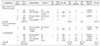
The 11 patients included 4 men and 7 women, with a mean age of 43.09±17.18 years (men, 55.25±13.55 years; women, 36.14±15.67 years). Dental treatments leading to emphysema included one case each of crown removal and subgingival curettage, and two cases each of tooth extraction, orthodontic miniscrew installation, class-V resin filling, tooth preparation, and root canal treatment. With regard to air distribution, there were five cases of subcutaneous emphysema within the facial area (45.5%; Group 1) and six cases of subcutaneous emphysema of the facial area spreading to the neck area and beyond (54.5%; Group 2). In Group 2, subcutaneous emphysema was localized to the neck in two cases (18.2%) and spread to the mediastinal region in the remaining four cases (36.4%). There was no significant difference in the duration of antibiotic use between Groups 1 and 2 (P=0.329). The mean length of follow-up was 1.8 weeks in both groups (P=0.931). Of the 11 patients, six were hospitalized (54.5%), with a mean duration of hospitalization of 4.80±1.10 days. Among these six patients, the subcutaneous emphysema spread to the neck and beyond in five cases, while in the remaining case the subcutaneous emphysema had spread extensively to the retropharyngeal space within the facial region. Of the 11 patients, two exhibited dyspnea (18.2%) in addition to subcutaneous emphysema, which spread to the mediastinum, including the neck region.(Table 1)
The results of our analysis of air-orifice locations revealed that subcutaneous emphysema originating from maxillary teeth (n=8; 66.7%) was two times more common than that originating from mandibular teeth (n=4; 33.3%). Subcutaneous emphysema originating from the posterior teeth (n=11; 91.7%) was also more common than that originating from the anterior teeth (n=1; 8.3%).(Table 2)
The results of our analysis of the range of emphysema according to the location of air orifices revealed that, among seven patients with subcutaneous emphysema introduced from the maxilla, only two presented with disease that had spread to the neck and beyond. On the other hand, all four patients with subcutaneous emphysema introduced from the mandible or both jaws exhibited disease that had spread to the neck and beyond. However, the difference in vertical location of air orifices (maxilla, mandible, or both; P=0.106) was not statistically significant. There was also no significant variation in the range of air distribution according to the anteroposterior location of air orifices (anterior teeth, premolars, or molars; P=0.545).(Table 3)
2. Major case
We report here the case of patient #7 as a representative of the study population. A 60-year-old woman with no medical history aside from thyroidectomy for thyroid cancer in 2012 was admitted to our institution with a chief complaint of swelling and discomfort in the right side of her face that had developed during prosthetic treatment at a private dental clinic on the same day as her admission. The event occurred during crown preparation of the right maxillary second premolar using a high-speed handpiece.
The vital signs of the patient at the time of admission were as follows: blood pressure, 162/93 mmHg; pulse, 93 beats/min; body temperature, 36.5℃; and respiratory rate, 20 breaths/min. Swelling was observed throughout the right orbital region, midface, and lower face. Crepitus could be felt by palpation from the right cheek to the supraclavicular region, and the patient complained of discomfort in these regions. Blood tests revealed a WBC count of 4,540/µL (normal range, 4,000-10,800/µL) with a normal neutrophil percentage (60.6%; normal range, 40%-73%) and count (2,750/µL; normal range, 2,000-7,000/µL).
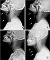
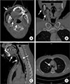
Although the patient showed no signs of dyspnea, neck x-ray radiographs showed extension of radiolucent layers up to the clavicular region.(Fig. 1. A) A head and neck CT scan showed multiple radiolucent regions with HU values (−943.8; mean HU of 3 arbitrary points) indicative of the presence of air throughout the right infraorbital, buccal, infratemporal, parapharyngeal, retropharyngeal, and submandibular spaces. (Fig. 2. A-C)
On the basis of her dental history as well as the radiographic findings and the results of palpation of the swollen areas demonstrating crepitus during clinical examination, the patient was diagnosed with subcutaneous emphysema secondary to her recent dental procedure. The subcutaneous emphysema had spread to the deep cervical region. Considering the risk of infection at the site of subcutaneous emphysema, the patient was hospitalized and administered conservative treatment by intravenous injection of prophylactic antibiotics (1 g cefotetan, two times a day).
Spread of subcutaneous emphysema to the mediastinal region was suspected and the Department of Cardiothoracic Surgery was thus consulted for confirmation and appropriate management with regard to the chest region. Chest CT scans showed small volumes of air distribution up to the mediastinum.(Fig. 2. D) The patient was administered conservative treatment with monitoring for saturation and oxygen supplementation by means of a nasal cannula.
After 4 days of hospitalization, the patient showed a decrease in swelling and loss of air layers on neck radiographs (Fig. 1. A-C) and was discharged. The patient was switched to oral antibiotics (100 mg cefdinir, three times a day) after discharge. As an outpatient at 6 days post-discharge, the patient showed complete resolution of both swelling and crepitus on palpation and absence of air on plain x-ray radiographs of the neck.(Fig. 1. D) Therefore, treatment and monitoring were considered to be complete.
IV. Discussion
1. Diagnosis
An appropriate differential diagnosis of subcutaneous emphysema and conditions that can present with head and neck swelling—such as infection, anaphylaxis, angioedema, and hematoma—is important7. Infectious diseases do not have a rapid enough onset to occur during or immediately after a dental procedure, patients must also have a suspected site of bacterial infection. Likewise, angioedema caused by an allergic reaction is commonly accompanied by swelling and itching. A major clinical characteristic of subcutaneous emphysema that differentiates it from other disease entities is the presence of crepitus on palpation of the swollen site217. In the present study, all 11 patients exhibited this symptom.
Patients with subcutaneous emphysema related to dental procedures usually have a history of dental procedures performed using a device that delivers high pressure, such as a dental handpiece or an air/water syringe13. Subcutaneous emphysema usually occurs during or immediately after a dental procedure2. Even when a high-pressure air device is not used, air can be introduced through disruption of the mucosal barrier at the wound or incision site18. For these reasons, careful dental history taking is important for diagnosing subcutaneous emphysema related to dental procedures. In addition, if subcutaneous emphysema has spread to the neck, radiolucent air layers may be observed on anteroposterior or lateral plain x-ray radiographs of the neck69 (Fig. 1. A), and multiple radiolucent bubble-like images can be observed on CT scans. (Fig. 2. A-C) Additionally, HU values of −1,000 (close to those of air) can be measured within the radiolucent regions on CT images1819. In the present study, all six patients with subcutaneous emphysema spreading to the neck region had undergone plain neck x-ray radiography, and five of these patients underwent CT imaging. All CT images exhibited HU values consistent with the presence of air.(Table 1)
2. Treatment and progress
Infection prophylaxis and airway management must be considered for the treatment of subcutaneous emphysema18. Because of the disruption of the oral mucosa, oral bacterial flora may be introduced through the air-inflow route, which can lead to cellulitis or abscess formation upon infection31215. Furthermore, infection at the potential site of subcutaneous emphysema can progress at a faster rate than common infections13. Therefore, patients must be administered conservative treatment with prophylactic antibiotics until the air is naturally absorbed and lost. As first-choice broad-spectrum antibiotics that can work against oral flora, penicillins are recommended for patients who are not allergic461120, and cephalosporins may also be used in this context21. Such antibiotics are usually orally administered, but intravenous administration after hospital admission is recommended if a significant risk of infection is anticipated because of the extensive spread of subcutaneous emphysema. Analgesics may be prescribed along with antibiotics for pain relief2.
Air is naturally absorbed and lost within soft tissue. Therefore, it is not necessary to perform additional incision and drainage for air removal. Indeed, unnecessary incision and drainage can create additional routes for air or bacterial inflow and induce inflammatory reactions through the new wound. Among all of the patients with subcutaneous emphysema in the present study, excluding those who underwent surgical extraction, patient #6 showed the greatest increase in WBC count and neutrophil ratio.(Table 1) In this case, subcutaneous emphysema that occurred after orthodontic miniscrew installation had been misdiagnosed as an abscess at a private dental clinic, and the patient had undergone unnecessary incision and drainage. Her condition worsened thereafter, and she subsequently visited the emergency room of our hospital.
In this study, patients were administered 100 mg cefdinir (every 8 hours, orally) and 1 g cefotetan (every 12 hours, intravenously) for antibiotic prophylaxis provided they were not allergic to these medications. Upon adequate conservative treatment with antibiotics, patients usually show spontaneous improvement within 3 to 5 days and complete recovery within 7 to 10 days13,14. In the present study, the average duration of antibiotic therapy was 8.55 days (standard deviation [SD], 4.46 days). Following an average of 1.82 weeks (SD, 1.19 weeks) of monitoring, all 11 patients showed improvement without any complications.
When subcutaneous emphysema spreads over large areas, it can spread to the parapharyngeal, retropharyngeal, and deep-neck space218 and cause dyspnea by compressing the airway. It can also spread to the mediastinum, which could affect respiratory or cardiac function2. Therefore, widespread subcutaneous emphysema requires hospitalization and patient monitoring. However, in the present study, there was no significant variation in the length of follow-up according to the range of air distribution in subcutaneous emphysema (P=0.931).(Table 1) This result may have been because subcutaneous emphysema is resolved not through inflammatory reactions but rather by means of air absorption within soft tissues. In contrast, abscesses take much longer to resolve depending on the extent of the infection.
If radiographic findings demonstrate spread of subcutaneous emphysema to the chest, clinicians should consider consultation with specialists in cardiothoracic surgery1822. Additionally, saturation monitoring is recommended if a patient suffers from dyspnea or is hospitalized because of widespread subcutaneous emphysema18. Oxygen inhalation through a cannula or mask can promote air absorption by reducing the partial pressure of N2 within blood, which can be beneficial for improvement23. In our study, of the six patients who presented with subcutaneous emphysema of the neck and beyond, four had subcutaneous emphysema spreading to the mediastinal area and were subsequently administered O2 therapy in cooperation with the Department of Cardiothoracic Surgery.(Table 2)
3. Patient education
To prevent additional air inflow, behaviors that increase intraoral pressure—such as forced exhalation, coughing, smoking, and gargling—must be avoided13. In addition, because lying in supine position can also worsen subcutaneous emphysema24, patients should be educated to position themselves with a high pillow or slightly raised bed in order to maintain their head above their chest.
4. Improvement evaluation
In this study, symptom improvement was determined by clinical examination for crepitus on palpation at the sites of subcutaneous emphysema. For subcutaneous emphysema spreading to the neck, it is useful to acquire new anteroposterior or lateral plain x-ray radiographs of the neck in order to evaluate for the loss of radiolucent air layers as an objective marker of improvement.(Fig. 1)
V. Conclusion
Subcutaneous emphysema encountered at the dental clinic can be easily diagnosed on the basis of palpable crepitus and history of dental treatment close to the time of onset. With appropriate conservative treatment administered along with antibiotic prophylaxis that considers the risk of infection, patients show improvement within a timeframe of a few days to weeks. In the present study population, there was no significant difference in treatment duration between patients with subcutaneous emphysema within the facial area and those with subcutaneous emphysema spreading to the neck and beyond. Going forward, it will be important to confirm these results in a larger population.
Hospitalization and thorough monitoring are recommended for patients with subcutaneous emphysema who present with widespread diffusion over the neck, difficulty in swallowing, or dyspnea. In cases where spread of disease to the mediastinal region is suspected, consultation with specialists in cardiothoracic surgery should be performed. Misdiagnosis and inappropriate treatment can lead rapid disease progression and infection or complications due to dyspnea. Thus, early diagnosis and accurate treatment of subcutaneous emphysema based on a thorough understanding of its characteristics are important.


 XML Download