

 PDF
PDF ePub
ePub Citation
 Print
Print


Kyoung Hee Kim1, Seung-Mi Jeong1 , Ye Chan Lee1, Xue Yin An1, Byung-Ho Choi2
, Ye Chan Lee1, Xue Yin An1, Byung-Ho Choi2
Abstract
In edentulous patients, implant - supported fixed prosthesis treatment has been proved to be useful, but involves complex treatment process. On the other hand, in the modern dentistry, digital technology has been developed day by day and it has expanded its range to the implant restoration of edentulous patients. In this case, a digital system was used for all stages of diagnosis, surgery, design and fabrication of provisional implants fixed prosthesis restoration in 66-year-old mandibular edentulous patients. In the preoperative diagnosis stage, a provisional restoration was designed based on the mucosal scan using the intraoral scanner and the stable occlusion of prefabricated complete denture of the patient. After flapless implant surgery using the surgical guide, the prefabricated interim restoration was connected to the implant and used as immediate provisional restoration. The final restoration was designed and fabricated by transferring the vertical dimension and the centric relation of the provisional restoration with stable occlusion using digital technology. We report a simple protocol of implant treatment in edentulous patients by using digital techniques to preserve the patient's vertical dimension and occlusion.
Figures and Tables
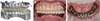
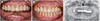 | Fig. 1Pre-operation. (A) Frontal view without existing denture, (B) Frontal view with existing denture, (C) Panoramic radiograph.
|
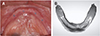 | Fig. 2Direct scan of residual ridge. (A) Marking on residual ridge using radiopaque resin, (B) 3D scan image of residual ridge.
|
 | Fig. 3Data of Occlusion. (A) Impression using denture, (B) Biting image, (C) Deletion of lower denture image.
|
 | Fig. 4Merging CT image with scan file and Planning of implant placement. (A) Right, (B) Front, (C) Left.
|
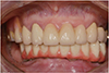
 | Fig. 6Design of interim prosthesis. (A) Frontal view, (B) Occlusion with antagonist jaw, (C) Occlusal view.
|
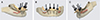
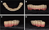
 | Fig. 7Pre-fabrication of interim prosthesis. (A) Occlusal view, (B) Frontal view, (C) Inner surface.
|
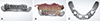
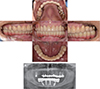
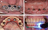 | Fig. 8Flapless Implantation and Interim prosthesis connection. (A) Surgical guide, (B) Implantation, (C) Confirming of cylinder site using interim prosthesis, (D) Adhesion of interim prosthesis and cylinder using resin curing.
|
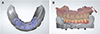 | Fig. 10Overlapping of scanfile of interim prosthesis and residual ridge. (A) Overlapping of scan files, (B) Confirming of implant site using interim prosthesis.
|
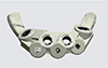
 | Fig. 11Fabrication of framework. (A) Design of framework using occlusion of provisional prosthesis, (B) Intraoral check of framework, (C) Checking of cleansing space using dental floss.
|
References
1. Wyatt CC. The effect of prosthodontic treatment on alveolar bone loss: a review of the literature. J Prosthet Dent. 1998; 80:362–366.

2. Kopp CD. Brånemark osseointegration. Prognosis and treatment rationale. Dent Clin North Am. 1989; 33:701–731.
3. Zarb GA, Schmitt A. The longitudinal clinical effectiveness of osseointegrated dental implants: the Toronto Study. Part II: The prosthetic results. J Prosthet Dent. 1990; 64:53–61.
4. Joda T, Brägger U. Digital vs. conventional implant prosthetic workflows: a cost/time analysis. Clin Oral Implants Res. 2015; 26:1430–1435.
5. Papaspyridakos P, Gallucci GO, Chen CJ, Hanssen S, Naert I, Vandenberghe B. Digital versus conventional implant impressions for edentulous patients: accuracy outcomes. Clin Oral Implants Res. 2016; 27:465–472.
6. Choi BH, Jeong SM. Digital flapless implantology. 1st ed. Seoul: Jisung Publishing;2015.
7. Tallgren A. The continuing reduction of the residual alveolar ridges in complete denture wearers: a mixed-longitudinal study covering 25 years. J Prosthet Dent. 1972; 27:120–132.
8. Zarb G, Bolender C. Prosthodontic treatment for edentulous patients. 12th ed. St. Louis: Mosby;2004.
9. Wismeijer D, van Waas MA, Kalk W. Factors to consider in selecting an occlusal concept for patients with implants in the edentulous mandible. J Prosthet Dent. 1995; 74:380–384.
10. Ting-Shu S, Jian S. Intraoral digital impression technique: A review. J Prosthodont. 2015; 24:313–321.
11. Ender A, Attin T, Mehl A. In vivo precision of conventional and digital methods of obtaining complete-arch dental impressions. J Prosthet Dent. 2016; 115:313–320.
12. Lee JH. Improved digital impressions of edentulous areas. J Prosthet Dent. 2017; 117:448–449.
 XML Download