

 PDF
PDF ePub
ePub Citation
 Print
Print


Abstract
To obtain denture retention, support, and stability in Class III edentulous cases with flat alveolar ridges and extensive flabby tissue is very difficult. The patient was a 72-year-old male who wore ill-fitting 20 year old dentures made by non-medical institutions. There was flabby tissue on the maxillary anterior ridge. The patient showed Angle Class III skeletal relationship with severe protruded mandible. First, temporary dentures were fabricated to restore the masticatory function, and final dentures were made through non- pressure impression technique and careful the arrangement of the posterior resin teeth. Improvement of the retention and stability of the denture during the occlusal force application is reported. (J Korean Acad Prosthodont 2018;56:295-301)
Go to : 
REFERENCES
1.Jacobson TE., Krol AJ. A contemporary review of the factors involved in complete denture retention, stability, and support. Part I: Retention. J Prosthet Dent. 1983. 49:5–15.

2.Atwood DA. Bone loss of edentulous alveolar ridges. J Peri-odontol. 1979. 50:11–21.
3.Klemetti E. A review of residual ridge resorption and bone density. J Prosthet Dent. 1996. 75:512–4.
4.Wical KE., Swoope CC. Studies of residual ridge resorption. I. Use of panoramic radiographs for evaluation and classification of mandibular resorption. J Prosthet Dent. 1974. 32:7–12.
5.Lynch CD., Allen PF. Management of the flabby ridge: using contemporary materials to solve an old problem. Br Dent J. 2006. 200:258–61.
6.Allen F. Management of the flabby ridge in complete denture construction. Dent Update. 2005. 32:524–6. 528.
7.Slade GD. Derivation and validation of a short-form oral health impact profile. Community Dent Oral Epidemiol. 1997. 25:284–90.
8.Ishikawa Y., Watanabe I., Hayakawa I., Minakuchi S., Uchida T. Evaluations of masticatory performance of complete denture wearers using color-changeable chewing gum and other evaluating methods. J Med Dent Sci. 2007. 54:65–70.
9.Phoenix RD., Engelmeier RL. Lingualized occlusion revisited. J Prosthet Dent. 2010. 104:342–6.
10.LaVere AM., Freda AL. Artificial tooth arrangement for prognathic patients. J Prosthet Dent. 1972. 28:650–4.
11.Gysi A. Special teeth for crossbite cases. Dent Digest. 1927. 33:167–71.
12.Sato Y., Ishida E., Minagi S., Akagawa Y., Tsuru H. The aspect of dietary intake of full denture wearers. Nihon Hotetsu Shika Gakkai Zasshi. 1988. 32:774–9.
13.Sanghvi SJ., Bhatt NA., Bhargava K. An evaluation of crossbite ridge relationships. A study of articulated jaw records of 150 edentulous patients. J Prosthet Dent. 1981. 45:24–9.
Go to :
 | Fig. 1.Pre-operative extraoral photographs. (A) Frontal view without denture, (B) Frontal view with denture, (C) Lateral view without denture, (D) Lateral view with denture. |

 | Fig. 3.Existing denture analysis. (A) Poor denture hygiene, (B, C, D) Comparison of existing denture with diagnostic study casts. |
 | Fig. 4.A severe interarch discrepancy due to skeletal class III relationship with mandibular prognathism was observed. (A) Lateral cephalometric radiograph, (B) Articulated study cast. |
 | Fig. 6.Test of mastication ability using color changeable chewing gum. (A) Before mastication, (B) Mastication result of existing denture, (C) Mastication result of temporary denture, (D) Color chart. |
 | Fig. 7.Maxillary final impression. (A) Impression taking, (B) Window opening of flabby tissue area, (C) Additional impression taking with light body silicon impression material, (D) Maxillary final impression. |
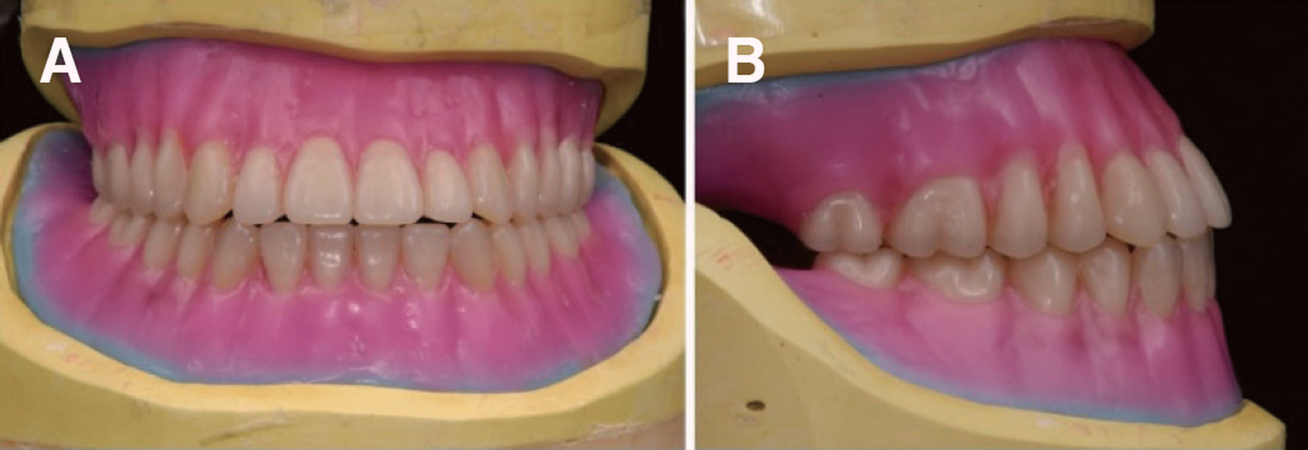
 | Fig. 8.Definitive denture tooth setup. (A, E) Semi-Anatomic, (B, D, F) Lingualized, (C, G, H) Cross bite. |
 | Fig. 9.Post insertion extraoral photographs. (A) Existing denture, Lateral view, (B) Provisional denture, Lateral view. |
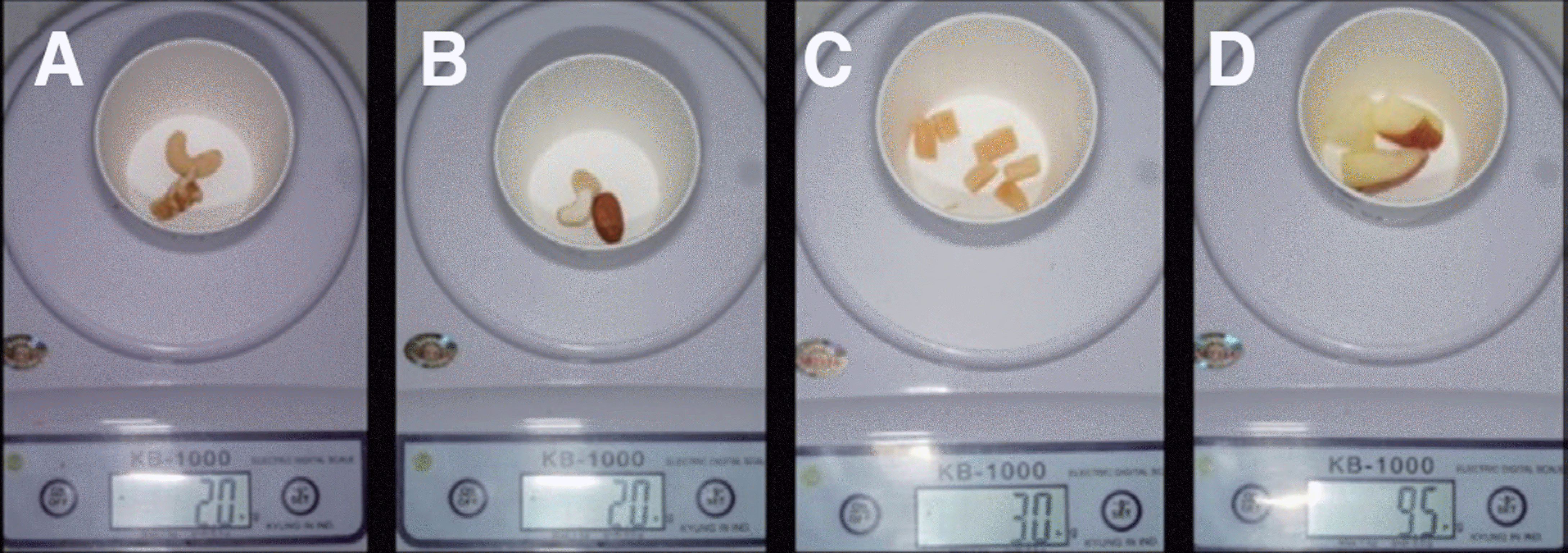 | Fig. 10.Food for masticatory test. (A) Cashew nut, walnut 2 g, (B) Cashew nut, Almond 2 g, (C) Semi-dried squid 3 g, (D) Apple 9.5 g. |
 | Fig. 11.Denture tilting test. (A) Cotton roll bite and impression taking with bite impression material, (B) Angle measurement. |
 XML Download