

 PDF
PDF ePub
ePub Citation
 Print
Print


Dear Editor:
Extramammary Paget's disease (EMPD) is a rare intraepidermal neoplastic disease that affects apocrine gland bearing skin such as scrotum, vulva, perineum, and axillae1. This disease arises in the cutaneous apocrine glands. Rarely, it also represents cutaneous extension from a visceral malignancy. In addition, several reports have suggested the association of Bowen's disease and squamous cell carcinoma in situ with EMPD23. One case of malignant melanoma (MM) coexistent with EMPD on the vulva at the same time has been reported previously4.
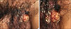
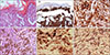
The study was approved by the Institutional Review Board of the St. Mary's Hospital, The Catholic University of Korea (IRB no. SC18ZESI0001). We received the patient's consent form about publishing all photographic materials. A 51-year-old man presented with seven years history of slowly growing non-healing nodules and plaques on the scrotum and pubic area. He had previously been treated with oral and topical antibiotics, antifungal agents, and topical steroid without improvement with the impression of tinea or eczema. There was also no history of internal malignancy. These lesions were not painful or pruritic. There were multiple, variable sized, and erythematous to skin colored plaques with black pigmented plaques and nodules at the base of his scrotum and pubic area (Fig. 1). Skin biopsy taken from the erythematous plaque showed epidermal infiltration by large atypical cells that had eosinophilic cytoplasm and prominent nuclei (Fig. 2A). Immunohistochemical staining showed that these tumor cells were strongly and diffusely positive for cytokeratin 7 (CK7), anti-human melanoma black 45 (Fig. 2B, C).
Skin biopsy taken from the blackish nodule showed nests of atypical melanocytes, presenting an infiltration in the direction of the dermis with poorly circumscribed and asymmetrical structural disturbance, usually marked nuclear atypia (Fig. 2D). Immunohistochemical staining showed that these tumor cells were strongly and diffusely positive for Melan A and S-100 (Fig. 2E, F).
We diagnosed this case as EMPD concurrent with MM of the same genital area based on clinical and histological examination results. The patient was transferred to a tertiary hospital for further evaluation and treatments.
EMPD is sometimes misdiagnosed as dermatitis, tinea cruris, candidiasis, psoriasis, contact dermatitis, and melanoma because EMPD presents as erythematous, scaly, and eczematous patches with plaques on the genital area. Histological differential diagnoses of perineal biopsies of this patient include squamous intraepithelial lesions, EMPD, and MM. These lesions can occur together, although this is rare2345. Histological findings of these diseases can overlap with each other2345. In such cases, immunohistochemical staining is helpful for differentiating EMPD from other diseases that also feature pagetoid spread. In general, EMPD is characterized by large and round cells with prominent central or peripheral nuclei and basophilic cytoplasm. It has disorganized intraepidermal proliferation of malignant epithelioid cells5. Immunohistochemical staining for CK7 is positive in tumor cells5. In our case, histopathologic feature of the nodule was presented with EMPD. However, the patch revealed a different feature, showing characteristics of MM.
The exact pathogenesis of EMPD remains unclear. Adnexal structures, pluripotent keratinocytes, and underlying internal organ malignancy may be involved. Similarly that of MM is also unanswered yet, but multifactorial interactions among genetic, host, and environmental factors has been involved in the development of MM4. These two diseases are not related each other in terms of pathogenesis, so this concurrence deserve to study throughly.
 XML Download