

 PDF
PDF ePub
ePub Citation
 Print
Print


Dear Editor:
Calcium and 1,25-dihydroxyvitamin D3 play important roles in epithelial cell functions. While low extracellular calcium levels in the culture medium induce rapid proliferation of keratinocytes, high extracellular calcium levels stimulate differentiation of keratinocytes. Vitamin D activates the proliferation of sebocytes and inhibits differentiation and sebum production in sebocytes1. In addition, vitamin D exerts an anti-inflammatory effect on sebocytes2. We evaluated the effects of extracellular calcium levels and the interaction between extracellular calcium levels and vitamin D on cultured human sebocytes.
Human sebocytes were cultured in Dulbecco's modified Eagle's medium (HyClone Laboratories, Logan, UT, USA) and Epilife (Gibco BRL, Grand Island, NY, USA) in a humidified atmosphere with 5% CO2 at 37℃. The sebocytes were used for the experiment after their second subculture. The sebocytes were cultured in the presence of higher extracellular calcium levels (0.25, 0.5, 0.75, 1, or 1.25 mM). CCK8 analysis revealed the absence of any toxic effect of extracellular calcium at the concentrations tested on cell viability. The active form of vitamin D 10−6 M was added into the culture media in the presence of each concentration of extracellular calcium. After the treatment of the sebocytes with vitamin D 10−6 M in the presence of 0.25, 0.5, 0.75, 1, or 1.25 mM extracellular calcium, real-time PCR analysis was performed in triplicate with a LightCycler (Roche Diagnostics, Indianapolis, IN, USA). The expression of biomarkers, such as interleukin (IL)-1β, IL-6, IL-8, and tumor necrosis factor (TNF)-α, in the sebocytes was measured in triplicate using ELISA (R&D Systems, Shanghai, China). Western blot analysis was performed to evaluate the expression of biomarkers, including psoriasin, LL-37, melanocortin-1 receptor (MC-1R), and MC-5R, in the sebocytes. Data were statistically analyzed with paired two-tailed Student's t-test using the statistical product and service solutions software (IBM Co., Armonk, NY, USA) for Windows (version 18). Data were presented as relative mean values±standard deviation. A value of p<0.05 was considered as statistically significant.
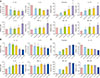
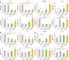
Higher extracellular calcium levels stimulated differentiation of sebocytes. The higher levels of extracellular calcium decreased the expression of IL-1β and IL-6 in gene and protein levels in the sebocytes (Fig. 1A, B). Gene and protein expression levels of IL-8, TNF-α, psoriasin, MC-1R, and MC-5R increased in the sebocytes in the presence of higher levels of extracellular calcium in a concentration-dependent manner (Fig. 1). In addition, higher extracellular calcium levels showed a concentration-dependent increase in the expression of LL-37 gene in the sebocytes (Fig. 1A). The treatment of the sebocytes with vitamin D resulted in a decrease in gene and protein expression of IL-1β (p<0.05), IL-6 (p<0.05), IL-8 (p<0.05), TNF-α, psoriasin (p<0.05) and MC-1R (Fig. 2). On the other hand, gene and protein expression of LL-37 (p< 0.05) and MC-5R in the sebocytes increased following their incubation with vitamin D (Fig. 2A, C). In the presence of higher concentrations of extracellular calcium, vitamin D treatment downregulated the expression of IL-1β (p<0.05), IL-6 (p<0.05), IL-8 and MC-1R (p<0.05), but not TNF-α (p<0.05) and psoriasin, in the sebocytes in gene and protein levels (Fig. 2). The treatment of the sebocytes with vitamin D in the presence of higher concentrations of extracellular calcium resulted in upregulation of LL-37, and MC-5R (p<0.05) expression in gene and protein levels (Fig. 2A, C).
The epidermal calcium gradient induces proliferation of basal keratinocytes, while it mediates differentiation of keratinocytes from the upper epidermal layers at higher extracellular calcium concentrations3. Higher extracellular calcium concentrations elevate the levels of intracellular free calcium. Vitamin D regulates cells expressing vitamin D receptors, such as keratinocytes. Some physiologic functions of vitamin D are associated with calcium. Vitamin D increases the intracellular calcium levels in keratinocytes, leading to their differentiation4. Therefore, both calcium and vitamin D can regulate the differentiation of epidermal keratinocytes. However, vitamin D inhibits the differentiation and sebum production in sebocytes5. Vitamin D may suppress sebocyte differentiation by binding to vitamin D receptor-retinoid X receptor dimers in a calcium-independent manner6. Like keratinocytes, higher extracellular calcium levels stimulated differentiation of cultured human sebocytes in this study. On the contrary, Zouboulis et al.7 reported that the increased extracellular calcium level stimulated proliferation of immortalized human SZ95 sebocytes but decreased extracellular calcium level induced differentiation of the sebocytes. Differentiation of sebocytes at higher extracellular calcium levels can be explained by the increase in the intracellular calcium levels and α-melanocyte-stimulating hormone (α-MSH)8. α-MSH not only increases intracellular calcium levels, but also stimulates MC-1R expression and lipid synthesis in immortalized sebocytes8. We also observed in this study that MC-1R and MC-5R were overexpressed in cultured human sebocytes at higher extracellular calcium levels. The differentiated sebocytes at higher extracellular calcium levels showed the increased expression of other biomarkers, such as IL-8, TNF-α, and psoriasin. As known previously, vitamin D inhibited the expression of IL-8, TNF-α, and psoriasin as well as IL-1β, IL-6, and MC-1R in cultured human sebocytes in this study. In addition, vitamin D inhibited the expression of IL-8 and MC-1R in cultured human sebocytes at higher extracellular calcium levels. However, the increased expression of TNF-α and psoriasin in the sebocytes was not inhibited by vitamin D. Like sebocytes in this study, keratinocytes has been known to induce psoriasin at higher calcium levels9. Furthermore, it was reported that TNF-α can be related to stimulate the expression of antimicrobial peptides10. In conclusion, extracellular calcium and vitamin D exert a regulatory function on the expression of biomarkers in cultured human sebocytes.
 XML Download