

 PDF
PDF ePub
ePub Citation
 Print
Print


Dear Editor:
Vitiligo is a common acquired depigmenting disorder of the skin and mucosa, affecting 0.5%~1% of the population worldwide1. It is classified into two major types, segmental and non-segmental, with the latter including several subtypes (generalized vitiligo, acrofacial vitiligo, and universal vitiligo). Segmental vitiligo (SV) is characterized by its early onset, rapid stabilization, and unilateral distribution1, whereas non-segmental vitiligo is often distributed symmetrically on the body and progresses slowly over time2. Currently, there is debate concerning whether the distribution patterns of SV, namely the dermatomal and Blaschko's linear distributions, indicate the disease origin.
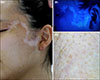
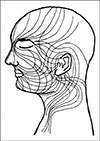
A 19-year-old female presented with a 2-year history of asymptomatic whitish patches, accompanied by poliosis, on her left temple. Following their sudden onset, the lesions showed no further development after a few months and, in contrast to dermatomes, formed an “S” shape (Fig. 1). The patient was diagnosed with SV along Blaschko's lines (Fig. 2)3. The study was approved by the Institutional Review Board of the Catholic Medical Center Office of Human Research Protection Program (VC17ZESE0096). We received the patient's consent form about publishing all photographic materials.
SV has been known to have a dermatomal distribution; however, in recent studies, the majority of SV lesions did not exactly fit this distribution pattern4. In one retrospective study, the distribution patterns of SV lesions were similar to those of certain mosaic skin disorders4. Furthermore, some authors have suggested that the recurrence pattern of SV corresponds well to cutaneous mosaicism1. The remarkable clinical similarity between SV and several cases of mosaic skin disorders involving melanocytes supports the hypothesis that cutaneous mosaicism is involved in SV4.
Although the pathogenesis of SV remains unclear, it has recently been hypothesized to include melanocyte mutations occurring during fetal development, a process known as somatic mosaicism that leads to a pigmented phenotype45. In this scenario, a single mutation in an emembryonic melanocyte would be passed on to its daughter cells, which later differentiate into functional melanocytes in the epidermis5. Somatic mutations in stress pathways may contribute to the unilateral distribution of SV; these abnormalities could result in localized autoimmunity in the area of altered melanocytes, while sparing normal cells located elsewhere5. Such a unilateral distribution of intrinsic melanocyte abnormalities would be distinct from that of dermatomes because the pathways of melanocyte migration are independent from cutaneous nerves4. Recently, increasing evidence of autoinflammation in SV has been published. One possible example of this phenomenon, termed “blaschkitis,” has been related to genetic mosaicism4. However, whether deregulation of the immune system is a casual factor of SV, or whether it arises secondary to cellular abnormalities in the epidermis, remains unclear4.
We herein presented a case of SV along Blaschko's lines, which implicates cutaneous mosaicism, rather than dermatomes, in the development of SV: further studies are needed to validate this hypothesis.
 XML Download