

 PDF
PDF ePub
ePub Citation
 Print
Print


INTRODUCTION
Morphea, also called localized scleroderma is a rare fibrosing disorder affecting the skin and underlying tissues. Segmental vitiligo is a distinct form of vitiligo, a common acquired pigmentation disorder, with dermatomal distribution or distribution of lesions along the Blaschko's lines. A few reports have previously noted the concurrent presentation of morphea and vitiligo at distinctly separate sites in the same patient12345678. However, it is extremely rare that these two diseases occur at the same site in a patient.
CASE REPORT
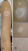
A 10-year-old Korean girl presented with multiple ill-defined hypopigmented patches on her left arm for 2 years (Fig. 1A). The lesions had an indurated, atrophic, and shiny area at the center. They gradually continued to increase in size over 2 years. Before presenting to outpatient clinic of Department of Dermatology, SMG-SNU Boramae Medical Center, she had received topical corticosteroids, excimer laser therapy, and Oriental remedies without any improvement. There was no history of trauma or injury to the site.
Physical examination revealed three depigmented patches with small satellite lesions on the left upper arm (Fig. 1A). The lesions were distributed along the T1 dermatome, and showed a central sclerotic area (Fig. 1B). In Wood's lamp examination, the lesions were accentuated compared to the peripheral uninvolved skin. The patient did not present with sclerodactyly, Raynaud's phenomenon, nailfold capillary changes, or signs of any systemic involvement, and a general physical examination was unremarkable.
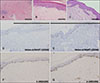
Punch biopsies performed at both sites, the hypopigmented area and normal uninvolved periphery revealed that the depigmented lesions demonstrated decreased basal pigmentation, mainly reduced numbers of melanocytes, as well as degenerated thickened dermal collagen bundles with focal dense lymphocytic infiltration, compared to adjacent normal skin (Fig. 2A, B). A biopsy from the normal periphery showed only mild superficial perivascular lymphocytic infiltration (Fig. 2C). Melan-A/MART-1 and S-100 positive melanocytes were noted to be decreased in the depigmented lesions compared to the normal periphery (Fig. 2D~G). Based on clinical and histopathological findings, she was diagnosed as circumscribed morphea accompanied by segmental vitiligo.
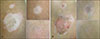
She was administered intra-lesional triamcinolone injections four times, and topical tacrolimus 0.1% ointment was prescribed to be applied twice daily. After six months of treatment, the lesions showed substantial improvement with repigmentation (Fig. 3).
We received the patient's consent about publishing all photographic materials.
DISCUSSION
A concurrent presentation of vitiligo and morphea has been reported, and such an association is noted to be more frequent than can be attributed to mere coincidence12345678. Based on some aspects of our case, we can propose that a definite relationship exists between the two diseases. In our patient, the two conditions progressed and improved simultaneously. Wood's lamp exam and results of specific stain (Melan-A/MART-1 and S-100 stain) favor vitiligo than a simple pigmentary change in morphea or lichen sclerosus. Also, the probability of scar tissue is low in that there was no traumatic history and previous performed oriental remedy was an oral medication that could not give trauma to the lesion. Furthermore, our case differed from previous reports in that the lesions of the two diseases were almost overlapping, and such an unique presentation supports our proposal that a definite association exists between the two diseases and their concurrent presentation is not a coincidence.
Various hypotheses have been proposed to explain the etiology of these two diseases; cutaneous mosaicism and autoimmunity are common to both conditions and may explain the presentation in our patient.
Interestingly, the lesions were distributed along Blaschko's lines in our case. Cutaneous mosaicism may explain the segmental distribution and linkage between the two diseases. A possible explanation could be that a clone of vulnerable cells is formed during embryogenesis and exposure of these cells to an appropriate trigger such as an autoimmune attack may result in the development of morphea and vitiligo9.
Morphea is considered to be an autoimmune disease based on various recent studies, and is associated with various other autoimmune diseases, that is, primary biliary cirrhosis10, myasthenia gravis11, Hashimoto's thyroiditis56, autoimmune thrombocytopenic purpura12, dermatomyositis, pemphigus13 and multiple autoimmune syndrome8. Moreover, circulating cytokines and soluble receptors are known to increase in patients with morphea; including interleukin-2 (IL-2), IL-4, IL-6, IL-13, soluble interleukin-2 (sIL-2) receptor and sIL-6, soluble CD4 and CD8, among others51314. Additionally, there is a higher incidence of autoantibodies such as anti-nuclear antibody (ANA) and anti-single-stranded DNA (ssDNA) in the serum of patients with morphea, and their relatives.51314. Vitiligo is also associated with various autoimmune diseases such as diabetes1516, pernicious anemia17, Addison's disease4, thyroid disease18, and alopecia areata4. Various organ-specific autoantibodies have been detected in the serum of patients with vitiligo515. In addition, the particular alleles of the multilocus major histocompatibility complex (MHC) are associated with susceptibility to vitiligo51920. Based on these findings, an autoimmune mechanism seems to be playing an important role in the concurrent presentation of the two diseases. This association therefore suggests that the regular screening for development of autoimmune diseases may be imperative in these patients.
Nevertheless, pathogenesis of morphea and vitiligo still eludes our understanding, the reported association between morphea and vitiligo, as was seen in our patient, does provide a better understanding of the pathogenesis of the two conditions. Further studies would be needed to elucidate whether the two conditions are definitively associated, and to determine a hypothesis that can explain the concurrence of the two diseases.
 XML Download