Abstract
Background
Citron is well known for an abundance of antioxidative and anti-inflammatory ingredients such as vitamin C, polyphenol compounds, flavonoids, and limonoids.
Objective
In this study, we aimed to evaluate the effects of citron essential oils on rosacea mediators in activated keratinocytes in vitro.
Methods
Normal human epidermal keratinocytes (NHEKs) were stimulated with 1α, 25-dihydroxyvitamin D3 (VD3) and interleukin 33 (IL-33) with LL-37 to induce rosacea mediators such as kallikrein 5 (KLK5), cathelicidin, vascular endothelial growth factor (VEGF), and transient receptor potential vanilloid 1 (TRPV1). These mediators were analyzed by performing reverse-transcription polymerase chain reaction (PCR), quantitative real-time PCR, immunocytofluorescence and enzyme-linked immunosorbent assay after NHEKs were treated with citron seed and unripe citron essential oils.
Citrus fruits are well known for their nutrition and health benefits. Yuzu (Citrus junos Tanaka), a common citrus fruit found in Korea and Japan, is rich in antioxidant phenols, and flavonoids with an anti-inflammatory action, such as limonene and hesperidin123. Hirota et al.4 showed that limonene extracted from citron reduced reactive oxygen species production and nuclear factor kappa-light-chain-enhancer of activated B cells (NF-kB) activity, indicating that citron has both antioxidant and anti-inflammatory properties4. However, despite the several beneficial effects of citron, no study has investigated its effects on the skin.
Rosacea is a common chronic inflammatory disease of the facial skin5; however, the exact pathogenesis of rosacea remains unclear. Recent studies have suggested that dysregulation of innate immunity and the neurovascular/neuroimmune system induces angiogenesis and inflammation, resulting in rosacea symptoms such as flushing, stinging, and telangiectasis678. Excessive production of LL-37 (an active peptide form of cathelicidin) and kallikrein 5 (KLK5), the predominant serine protease responsible for cleavage of cathelicidin into LL-37, has been reported to play a role in the dysregulation of innate immunity91011. LL-37 production is induced by the vitamin D pathway in human keratinocytes; therefore, ultra violet light acts as a trigger for rosacea12. Transient receptor potential vanilloid 1 (TRPV1), also known as the capsaicin receptor, has been reported to be involved in neurogenic dysregulation. It can be activated by heat, ethanol, or spicy food, all of which are triggers for rosacea131415. Vascular endothelial growth factor (VEGF), which has been reported to enhance angiogenesis and/or lymphangiogenesis in the skin with rosacea lesions, is associated with neurovascular dysfunction1617. Interleukin 33 (IL-33), a pro-inflammatory cytokine and an inducer of Th2-mediated responses, has been reported to enhance VEGF expression in keratinocytes18. In addition, abnormally increased expression of LL-37 may induce VEGF expression in epidermal keratinocytes, which corresponds to the enhanced inflammation and vascular response seen in rosacea19.
Therefore, LL-37, KLK5, TRPV1, and VEGF are considered as therapeutic targets for rosacea. This study was aimed at determining the effects of citron essential oils at different concentrations on rosacea mediators, KLK5, LL-37 (induced by 1α, 25-dihydroxyvitamin D3 [VD3]), VEGF, and TRPV1 induced by IL-33 and LL-37 in normal human epidermal keratinocytes (NHEKs) in vitro and evaluating their potential as adjuvant therapeutic agents for rosacea.
Two types of citron essential oils were used in this study, citron seed essential oil and unripe citron essential oil. Citrons were obtained from Goheung, Jeollanam-do. Citron essential oils were extracted by Hisol Co., Ltd (Namwon, Korea).
NHEKs were purchased from EpiLife (Cascade Biologics, Portland, OR, USA). The cells were cultured in basal keratinocyte growth media (EpiLife) supplemented with human keratinocyte growth supplement and antibiotics (100 U/ml penicillin and 100 µg/ml streptomycin) in a 5% CO2 incubator. Passages 2–9 were used for all experiments. NHEKs (1×10⁴ cells/well) were seeded in a 96-well plate. After the cells were treated with various concentrations of the essential oils, cell viability was determined using the 3-(4,5-dimethylthiazol-2-yl)-2,5-diphenyltetrazolium bromide (MTT; Sigma-Aldrich, St. Louis, MO, USA) assay according to the manufacturer's instructions20.
To induce KLK and LL-37 expression, NHEKs at 70% confluence were stimulated with VD3 (Sigma-Aldrich)2122. The expression of KLK5, LL-37, and vitamin D receptor (VDR) was evaluated at times ranging from 0 to 48 h and doses ranging from 10−9 to 10−7 M (1 to 100 nM)2324. Regulation of KLK 5, LL-37, and VDR induced by VD3 was also evaluated after the cells were treated with citron essential oils at various concentrations (0.005%, 0.01%, and 0.02%).
NHEKs at 70% confluence were exposed to different concentrations of recombinant human IL-33 (100 ng/ml; MACS, Auburn, CA, USA) and LL-37 peptides (0.5 µg/ml; ANYGEN, Jangseong, Korea). The expression of VEGF and TRPV1 was evaluated at times ranging from 0 to 48 h and doses ranging from 0.1 to 10 ng/ml19. Regulation of VEGF and TRPV1 was also evaluated after the cells were treated with citron essential oils at various concentrations (0.005%, 0.01%, and 0.02%).
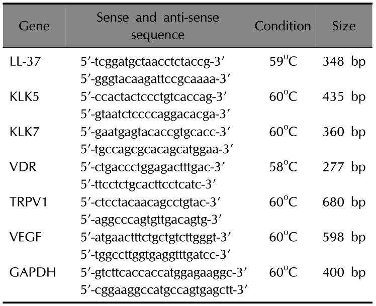
Total messenger RNA (mRNA) was isolated from human keratinocytes by using the RNeasy mini kit (Qiagen, Valencia, CA, USA). cDNA was synthesized using the Omniscript RT kit (Qiagen): 1 µg RNA, 10× buffer, dNTP, oligodT, inhibitor, reverse transcriptase, at 37℃ for 1 h, 93℃ for 5 min. A PCR was performed using the PCR-premixture kit (ELPIS, Daejeon, Korea) according to the manufacturer's instructions. Table 1 shows the primer sequences, PCR conditions, and product sizes. The PCR products were analyzed using 1.5% agarose gel electrophoresis, stained with Sybr Safe DNA gel stain buffer (Invitrogen, Carlsbad, CA, USA), and visualized using a luminescent image analyzer (LAS 3000; Fujifilm, Tokyo, Japan).
Total mRNA was isolated using an RNeasy mini kit (Qiagen). Subsequently, cDNA was reverse transcribed from 500 ng of total RNA with the Omniscript RT kit (Qiagen) and subjected to PCR with the HiPi PCR PreMix (ELPIS). The expression levels were normalized to endogenous glyceraldehyde 3-phosphate dehydrogenase (GAPDH) levels. To determine mRNA levels, RT-PCR and quantitative real-time PCR were performed with the same primer sets for the target genes. Real-time PCR was performed in triplicate with the HOT FIREPol EvaGreen® qPCR Mix Plus (Solis BioDyne, Tartu, Estonia) using a RotorGene 3000 system (Corbett Research, Cambridge, UK). The thermal cycling conditions were as follows: 15 min at 95℃, followed by 40 cycles at 95℃ for 10 s, 55℃–60℃ for 20 s, and 72℃ for 30 s. The relative abundance of a given transcript was estimated using the 2−ΔΔCt method, following normalization to GAPDH levels.
The cell suspensions were fixed with paraformaldehyde, blocked with 5% goat serum, and incubated with rabbit polyclonal anti-cathelicidin (LL-37) antibody (Abcam, Cambridge, MA USA), mouse monoclonal anti-VEGF antibody (Novusbio Biological, Littleton, CO, USA), or rabbit polyclonal anti-VR1 (TRPV1) antibody (Abcam). AlexFluor 488-conjugated goat anti-rabbit immunoglobulin (Ig) G and AlexaFluor 594-conjugated goat anti-mouse IgG (Molecular Probes; Invitrogen, Carlsbad, CA, USA) were used as secondary antibodies. Sections were mounted using 4′,6-diamidino-2-phenylindole (DAPI; Vector Laboratories, Burlingame, CA, USA). The images were visualized using confocal microscopy with a laser scanning microscope (LSM 510; Carl Zeiss, Jena, Germany) and analyzed using the LSM 5 browser imaging software.
Commercial ELISA kits were used according to the manufacturers' protocols to quantify the immune molecules of interest: human LL37 (Hycultbiotech, Uden, Netherlands), human KLK5 (R&D Systems, Inc., Minneapolis, MN, USA), human VEGF (R&D Systems, Inc.), and human TRPV1 (Mybiosource, San Diego, CA, USA).
All values are expressed as the mean±standard deviation. Statistical analyses were performed using one-way analysis of variance with a post hoc least significant difference test using IBM SPSS Statistics version 21.0 (IBM Co., Armonk, NY, USA) for multiple comparisons. A p-value less than 0.05 was considered statistically significant.
Cell viability was evaluated after the cells were exposed to various concentrations of citron seed essential oil and unripe citron essential oil (0, 0.001%, 0.002%, 0.004%, 0.008%, 0.016%, 0.031%, 0.063%, 0.125%, 0.25%, 0.5%, and 1%). Viability decreased upon treatment with 1% citron seed essential oil and 0.125% unripe citron essential oil (Supplementary Fig. 1).
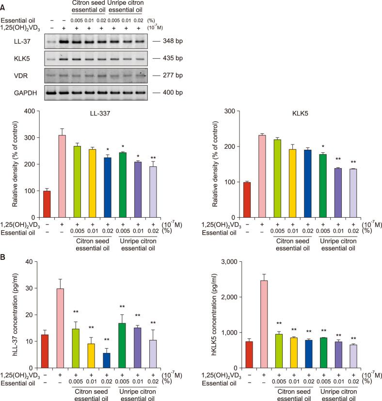
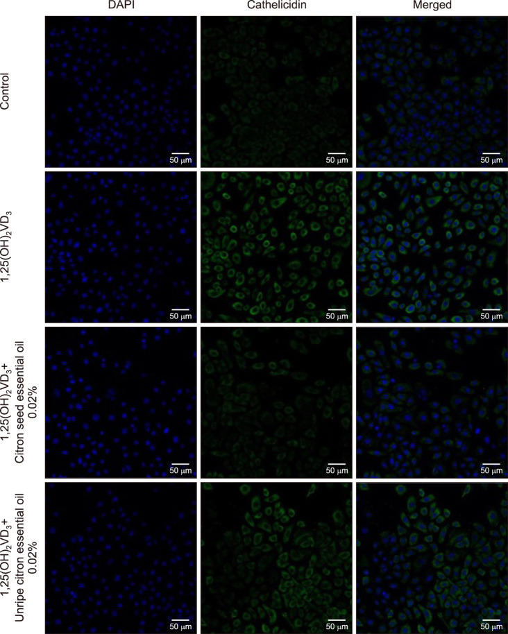
Stimulation of NHEKs with VD3 at a dose of 10−7 M for 24 h increased the levels of KLK5 and LL-37 maximally. After treatment of NHEKs with citron essential oils, KLK5, LL-37 and VDR mRNA expression induced by VD3 decreased. The mRNA levels of LL37 decreased after treatment with citron seed (0.02%) and unripe citron essential oils (≥0.005%), partially showing a dose-dependent pattern (Fig. 1A). Furthermore, the mRNA levels of KLK5 decreased after treatment with unripe citron essential oil (≥0.005%). Citron seed essential oil also decreased the mRNA level of KLK5, but the difference was not significant. Treatment with 0.02% unripe citron essential oil caused the maximum suppression of LL-37 and KLK5 mRNA levels. Further, ELISA showed that protein levels of LL-37 and KLK5 significantly decreased after treatment with citron essential oils at any concentration (Fig. 1B). Immunocytofluorescence showed that LL37 expression induced by VD3 decreased after treatment with 0.02% citron essential oils; this finding was consistent with previous results (Fig. 2).
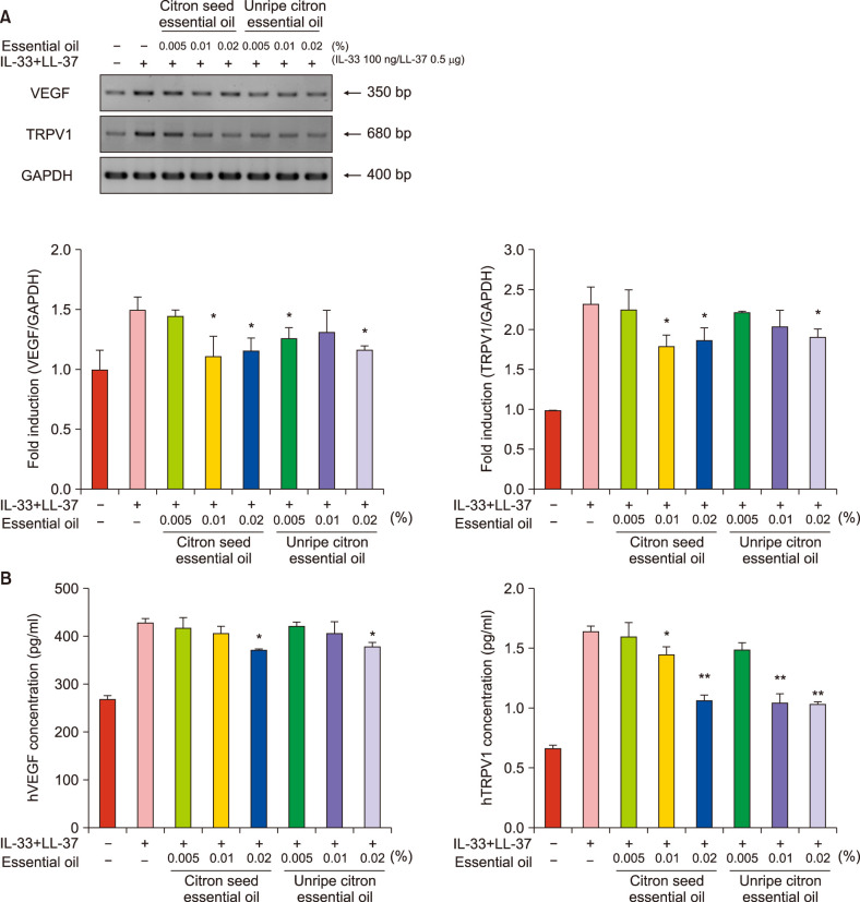
The levels of VEGF and TRPV1 maximally increased upon NHEK exposure to IL-33 100 ng/LL-37 0.5 µg for 24 h. After treatment with citron essential oils, the mRNA levels of VEGF and TRPV1 decreased and the extent of decrease was significant: citron seed essential oil, VEGF ≥0.01% and TRPV1 ≥0.01%, and unripe citron essential oil, VEGF 0.005% and 0.02%, and TRPV1 0.02% (Fig. 3A). Further, ELISA showed that protein levels of VEGF and TRPV1 significantly decreased after treatment with citron seed essential oil (VEGF and TRPV1: 0.02%) and unripe citron essential oil (VEGF and TRPV1: ≥0.01%) (Fig. 3B). Immunocytofluorescence showed that the IL-33-induced TRPV1 and VEGF expression in NHEKs decreased after treatment with 0.02% citron essential oil; this finding was consistent with previous results (Supplementary Fig. 2).
Citron is popular for its health benefits. Recent studies suggest that these various pharmacological effects that inhibit disease are known as antioxidant vitamin C and E, polyphenols and flavonoids that are found in many fruits2526. Naringin, a flavonoid present in many citrus fruits including citron, is non-toxic to humans, has a cholesterol-lowering effect, transforms leukemia cells into normal cells, inhibits the proliferation of breast cancer cells, which are known as anti-inflammatory and natural antimicrobial agents2728. Another flavonoid present in citron, hesperidin, is also known to have beneficial effects, such as antioxidant, hypotensive, and antiallergic effects. It is present in a large amount in citrus peels and inhibits an increase in blood cholesterol concentration29. In addition, citron is rich in limonoids such as limonin and normalin, which are excellent antioxidants3031. Limonin is one of the main components that lend bitterness to citrus fruits32. It has been reported to have antibacterial, antiviral, antinociceptive, anti-inflammatory, and anticarcinogenic effects333435. Further, although citron seeds account for 14% to 16% of the total citron fresh weight and most citron seeds are collected and discarded, they are richer in limonoids than is the whole fruit303136. In our previous experiments, we analyzed the effect of citron essential oils and five other essential oils (lavender, rosemary, remongrass, chamomile, and peppermint oil) on NHEKs activated by VD3. RT-PCR and real-time PCR showed that citron essential oils were effective in inhibiting LL-37 and KLK5 (Supplementary Fig. 3). Our additional analyses showed that citron essential oils are abundant in polyphenol (0.5 mg gallic acid equivalents [GAE]/kg in citron seed essential oil and 3.8 mg GAE/kg in unripe citron essential oil). This finding was consistent with those reported previously. Therefore, in this study, we used citron seed and unripe citron essential oils.
This study demonstrates that citron essential oils have a suppressive effect on LL-37, KLK5, TRPV1, and VEGF, which are the main components involved in the pathophysiology of rosacea, in vitro. Augmentation of the cathelicidin innate immune pathway (specifically KLK5 and LL-37) has been reported to be a major contributor to the pathophysiology of rosacea. Rosacea patients have an increased baseline expression of KLK5 and LL-37. KLK5 is the major serine protease responsible for cleaving cathelicidin into its active form, LL-37910. In rosacea-affected skin, LL-37 is processed into shorter fragments that regulate processes such as leukocyte chemotaxis, angiogenesis, and expression of extracellular matrix components9113738. Therefore, agents suppressing the production of KLK5 and thereby inhibiting LL-37 could markedly affect inflammation associated with rosacea. In this study, citron essential oils suppressed the levels of LL37 and KLK5 induced by VD3 in NHEKs. These results support that citron essential oils may normalize the dysregulation of the innate immune system in the skin of rosacea patients and help prevent and improve the symptoms of rosacea.
Recently, TRPV1 and TRP ankyrin receptor (TRPA1) have been found to contribute to the pathophysiology of rosacea. TRPV1 is expressed by sensory nerves and other non-neural cells, such as keratinocytes, and plays a role in vasoregulation and nociception1339. Although the signaling pathways of these receptors are not completely understood, they are activated by rosacea trigger factors such as heat, ethanol, and spicy food40. These receptors release important neuropeptides in neurogenic inflammation, such as substance P and calcitonin gene-related peptide41. In patients with rosacea, these receptors are upregulated, resulting in neurogenic dysregulation leading to rosacea symptoms such as persistent erythema and inflammation838. Although the role of angiogenesis in rosacea is controversial, some studies the support role of angiogenesis in rosacea pathophysiology617. Increased VEGF levels have been reported in lesional skin or rosacea, which result in enhanced inflammation and vascular response seen in rosacea1617. In this study, the levels of VEGF and TRPV1 induced by IL-33 in NHEKs decreased after treatment with citron essential oils. These results demonstrate that citron essential oils may improve rosacea symptoms such as flushing and telangiectasia by suppressing neurogenic dysregulation and angiogenesis in the skin of rosacea patients.
ELISA and immunocytofluorescence revealed that citron seed essential oil had a better inhibitory effect on KLK5 induced by VD3 than did unripe citron essential oil. Furthermore, RT-PCR showed that the inhibitory effects of citron seed essential oil on VEGF and TRPV1 were greater than those of unripe citron essential oil, but the difference was minimal and a dose-dependent pattern was not observed. Thus, the two oils showed comparable results in our experiments. Unlike unripe citron, the citron seed is abandoned in the manufacturing processes. Therefore, it is expected that citron seed will be useful economically and environmentally for development of therapeutic agents for rosacea.
There are several limitations to this study. First, no analysis was performed to determine which components of citron essential oils inhibited the rosacea-related factors. In addition, the mechanisms underlying the inhibitory actions remain to be elucidated, and further experiments are required. To address these limitations, an analysis of the ingredients of citron essential oils and additional experiments are underway.
In this study, it was demonstrated that citron seed and unripe citron essential oils suppressed LL-37, KLK5, VEGF, and TRPV1 in NHEKs stimulated with VD3 and IL-33. LL-37, KLK5, VEGF, and TRPV1 are mediators that play important roles in the pathophysiology of rosacea; hence, citron essential oils are expected to improve rosacea symptoms by inhibiting the underlying pathomechanism. On the basis of our results, we anticipate that citron essential oils could be valuable ingredients for an adjuvant therapeutic agent for rosacea.
ACKNOWLEDGMENT
This research was supported by the Ministry of Trade, Industry & Energy (MOTIE), Korea Institute for Advancement of Technology (KIAT) through the Encouragement Program for The Industries of Economic Cooperation Region.
References
1. Sawamura M, Wu Y, Fujiwara C, Urushibata M. Inhibitory effect of yuzu essential oil on the formation of N-nitrosodimethylamine in vegetables. J Agric Food Chem. 2005; 53:4281–4287. PMID: 15884872.
2. Yoo KM, Lee KW, Park JB, Lee HJ, Hwang IK. Variation in major antioxidants and total antioxidant activity of Yuzu (Citrus junos Sieb ex Tanaka) during maturation and between cultivars. J Agric Food Chem. 2004; 52:5907–5913. PMID: 15366841.
3. Zou Z, Xi W, Hu Y, Nie C, Zhou Z. Antioxidant activity of citrus fruits. Food Chem. 2016; 196:885–896. PMID: 26593569.
4. Hirota R, Roger NN, Nakamura H, Song HS, Sawamura M, Suganuma N. Anti-inflammatory Effects of limonene from yuzu (Citrus junos Tanaka) essential oil on eosinophils. J Food Sci. 2010; 75:H87–H92. PMID: 20492298.
5. Crawford GH, Pelle MT, James WD. Rosacea: I. Etiology, pathogenesis, and subtype classification. J Am Acad Dermatol. 2004; 51:327–341. PMID: 15337973.
6. Schwab VD, Sulk M, Seeliger S, Nowak P, Aubert J, Mess C, et al. Neurovascular and neuroimmune aspects in the pathophysiology of rosacea. J Investig Dermatol Symp Proc. 2011; 15:53–62.
7. Two AM, Del Rosso JQ. Kallikrein 5-mediated inflammation in rosacea: clinically relevant correlations with acute and chronic manifestations in rosacea and how individual treatments may provide therapeutic benefit. J Clin Aesthet Dermatol. 2014; 7:20–25.
8. Steinhoff M, Schauber J, Leyden JJ. New insights into rosacea pathophysiology: a review of recent findings. J Am Acad Dermatol. 2013; 69:S15–S26. PMID: 24229632.
9. Yamasaki K, Di Nardo A, Bardan A, Murakami M, Ohtake T, Coda A, et al. Increased serine protease activity and cathelicidin promotes skin inflammation in rosacea. Nat Med. 2007; 13:975–980. PMID: 17676051.
10. Yamasaki K, Schauber J, Coda A, Lin H, Dorschner RA, Schechter NM, et al. Kallikrein-mediated proteolysis regulates the antimicrobial effects of cathelicidins in skin. FASEB J. 2006; 20:2068–2080. PMID: 17012259.
11. Morizane S, Yamasaki K, Mühleisen B, Kotol PF, Murakami M, Aoyama Y, et al. Cathelicidin antimicrobial peptide LL-37 in psoriasis enables keratinocyte reactivity against TLR9 ligands. J Invest Dermatol. 2012; 132:135–143. PMID: 21850017.
12. Schauber J, Gallo RL. The vitamin D pathway: a new target for control of the skin's immune response? Exp Dermatol. 2008; 17:633–639. PMID: 18573153.
13. Pecze L, Szabó K, Széll M, Jósvay K, Kaszás K, Kúsz E, et al. Human keratinocytes are vanilloid resistant. PLoS One. 2008; 3:e3419. PMID: 18852901.
14. Caterina MJ, Schumacher MA, Tominaga M, Rosen TA, Levine JD, Julius D. The capsaicin receptor: a heat-activated ion channel in the pain pathway. Nature. 1997; 389:816–824. PMID: 9349813.
15. Sulk M, Seeliger S, Aubert J, Schwab VD, Cevikbas F, Rivier M, et al. Distribution and expression of non-neuronal transient receptor potential (TRPV) ion channels in rosacea. J Invest Dermatol. 2012; 132:1253–1262. PMID: 22189789.
16. Huggenberger R, Detmar M. The cutaneous vascular system in chronic skin inflammation. J Investig Dermatol Symp Proc. 2011; 15:24–32.
17. Gomaa AH, Yaar M, Eyada MM, Bhawan J. Lymphangiogenesis and angiogenesis in non-phymatous rosacea. J Cutan Pathol. 2007; 34:748–753. PMID: 17880579.
18. Balato A, Lembo S, Mattii M, Schiattarella M, Marino R, De Paulis A, et al. IL-33 is secreted by psoriatic keratinocytes and induces pro-inflammatory cytokines via keratinocyte and mast cell activation. Exp Dermatol. 2012; 21:892–894. PMID: 23163661.
19. Borelli C, Becker B, Thude S, Fehrenbacher B, Isermann D. Dermasence refining gel modulates pathogenetic factors of rosacea in vitro. J Cosmet Dermatol. 2017; 16:e31–e36. PMID: 28349651.
20. Denizot F, Lang R. Rapid colorimetric assay for cell growth and survival: modifications to the tetrazolium dye procedure giving improved sensitivity and reliability. J Immunol Methods. 1986; 89:271–277. PMID: 3486233.
21. Schauber J, Dorschner RA, Coda AB, Büchau AS, Liu PT, Kiken D, et al. Injury enhances TLR2 function and antimicrobial peptide expression through a vitamin D–dependent mechanism. J Clin Invest. 2007; 117:803–811. PMID: 17290304.
22. Thibaut de Ménonville ST, Rosignoli C, Soares E, Roquet M, Bertino B, Chappuis JP, et al. Topical treatment of rosacea with ivermectin inhibits gene expression of cathelicidin innate immune mediators, LL-37 and KLK5, in reconstructed and ex vivo skin models. Dermatol Ther (Heidelb). 2017; 7:213–225. PMID: 28243927.
23. Lee JB, Bae SH, Moon KR, Na EY, Yun SJ, Lee SC. Light-emitting diodes downregulate cathelicidin, kallikrein and toll-like receptor 2 expressions in keratinocytes and rosacea-like mouse skin. Exp Dermatol. 2016; 25:956–961. PMID: 27315464.
24. Morizane S, Yamasaki K, Kabigting FD, Gallo RL. Kallikrein expression and cathelicidin processing are independently controlled in keratinocytes by calcium, vitamin D(3), and retinoic acid. J Invest Dermatol. 2010; 130:1297–1306. PMID: 20090765.
25. Hertog MG, Feskens EJ, Hollman PC, Katan MB, Kromhout D. Dietary antioxidant flavonoids and risk of coronary heart disease: the Zutphen Elderly Study. Lancet. 1993; 342:1007–1011. PMID: 8105262.
26. Vinson JA, Su X, Zubik L, Bose P. Phenol antioxidant quantity and quality in foods: fruits. J Agric Food Chem. 2001; 49:5315–5321. PMID: 11714322.
27. Lee SL, Seo CS, Kim JH, Shin HK. Contents of poncirin and naringin in fruit of poncirus trifoliata according to different harvesting times and locations for two years. Korean J Pharmacogn. 2011; 42:138–143.
28. Yang HS, Eun JB. Fermentation and sensory characteristics of Korean traditional fermented liquor (makgeolli) added with citron (Citrus junos SIEB ex TANAKA) juice. Korean J Food Sci Technol. 2011; 43:438–445.
29. Woo DH. Stabilization to sunlight of natural coloring matter by soluble methyl-hesperidin. Korean J Food Sci Technol. 2000; 32:50–55.
30. Kim SY, Shin KS. Evaluation of physiological activities of the citron (Citrus junos Sieb. ex TANAKA) seed extracts. Prev Nutr Food Sci. 2013; 18:196–202. PMID: 24471132.
31. Woo KL, Kim JI, Kim MC, Chang DK. Determination of flavonoid and limonoid compounds in citron (Citrus junos Sieb. et Tanaka) seeds by HPLC and HPLC/MS. J Korean Soc Food Sci Nutr. 2006; 35:353–358.
32. Manners GD. Citrus limonoids: analysis, bioactivity, and biomedical prospects. J Agric Food Chem. 2007; 55:8285–8294. PMID: 17892257.
33. Miller EG, Gonzales-Sanders AP, Couvillon AM, Wright JM, Hasegawa S, Lam LK. Inhibition of hamster buccal pouch carcinogenesis by limonin 17-beta-D-glucopyranoside. Nutr Cancer. 1992; 17:1–7. PMID: 1574440.
34. El-Readi MZ, Hamdan D, Farrag N, El-Shazly A, Wink M. Inhibition of P-glycoprotein activity by limonin and other secondary metabolites from citrus species in human colon and leukaemia cell lines. Eur J Pharmacol. 2010; 626:139–145. PMID: 19782062.
35. Langeswaran K, Jagadeesan AJ, Revathy R, Balasubramanian MP. Chemotherapeutic efficacy of limonin against Aflatoxin B1 induced primary hepatocarcinogenesis in Wistar albino rats. Biomed Aging Pathol. 2012; 2:206–211.
36. Giamperi L, Fraternale D, Bucchini A, Ricci D. Antioxidant activity of citrus paradisi seeds glyceric extract. Fitoterapia. 2004; 75:221–224. PMID: 15030930.
37. Koczulla R, von Degenfeld G, Kupatt C, Krötz F, Zahler S, Gloe T, et al. An angiogenic role for the human peptide antibiotic LL-37/hCAP-18. J Clin Invest. 2003; 111:1665–1672. PMID: 12782669.
38. Two AM, Wu W, Gallo RL, Hata TR. Rosacea: part I. Introduction, categorization, histology, pathogenesis, and risk factors. J Am Acad Dermatol. 2015; 72:749–758. PMID: 25890455.
39. Earley S. Vanilloid and melastatin transient receptor potential channels in vascular smooth muscle. Microcirculation. 2010; 17:237–249. PMID: 20536737.
40. Wang TT, Nestel FP, Bourdeau V, Nagai Y, Wang Q, Liao J, et al. Cutting edge: 1,25-dihydroxyvitamin D3 is a direct inducer of antimicrobial peptide gene expression. J Immunol. 2004; 173:2909–2912. PMID: 15322146.
41. Nilius B, Owsianik G, Voets T, Peters JA. Transient receptor potential cation channels in disease. Physiol Rev. 2007; 87:165–217. PMID: 17237345.
SUPPLEMENTARY MATERIALS
Supplementary data can be found via http://anndermatol.org/src/sm/ad-30-653-s001.pdf.