

 PDF
PDF ePub
ePub Citation
 Print
Print


I. Introduction
Ameloblastoma is a benign odontogenic tumor that often shows aggressive growth and has a high rate of recurrence1. It is an aggressive benign tumor of epithelial origin that may arise from the enamel, remnants of the dental lamina, the lining of odontogenic cysts, or possibly from the basal epithelial cells of the oral mucosa2. The treatment of ameloblastoma varies based on the clinical, histopathologic, and radiographic characteristics. Unicystic forms are thought to be less likely to recur; however, although a more conservative approach is recommended, it is seldom adopted in practice3. Typically, aggressive surgical treatments, such as marginal or segmental resection, have been implemented, but some conservative surgical methods are also being introduced, including decompression, enucleation, or curettage45. This case series show the potential for favorable healing without recurrence in ameloblastoma patients treated with conservative surgery.
II. Cases Report
1. Case 1
In 2015, a 24-year-old male was referred to our hospital for evaluation of gingival swelling in the mandibular left posterior region. The patient was a non-smoker and had no underlying disease. He had first noticed the lesion about one year prior. On extra-oral examination, facial asymmetry with swelling involving the left side of the mandible was observed at the time of admission. Intraoral examination showed a discharge of pus in the posterior region.
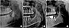
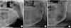
X-ray radiographs (panoramic radiograph and computed tomography [CT]) were performed.(Fig. 1. A) Axial CT showed a fairly large (27 mm×34 mm×23 mm) and well-defined rounded cystic destructive lesion involving the left mandibular body. Absorption of the roots of #36 and #37 had occurred. Incisional biopsy was performed under local anesthesia, and the tissue was sent for histopathological examination, which revealed a suggestive benign cyst. Endodontic treatment of #36 was performed.
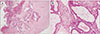
At the time of operation, extraction of #37 and #38 and enucleation of the lesion were carried out under general anesthesia. The iliac bone graft took place at the same time. After surgery, a newly excisional biopsy was performed, and the results did not show a benign cyst but instead were suggestive of unicystic ameloblastoma, mural type.(Fig. 2) A follow-up check was done at three months, six months, nine months, and one year after surgery. Nine months postoperatively, a panoramic radiograph revealed favorable healing of the iliac bone graft in the left mandibular body.(Fig. 1. B)
At one year and five months after surgery, implantation of #37 was performed. The implantation was done using a one-stage technique because the initial torque was favorable. (Fig. 1. C) Based on the results of the initial histopathological examination, cyst enucleation and an iliac bone graft were performed simultaneously in this patient. Usually in cases of ameloblastoma, a bone graft is not carried out at the same time due to the high recurrence rate. In this case, non-invasive cyst enucleation and the iliac bone graft were performed simultaneously, and the healing was satisfactory without any evidence of recurrence.
2. Case 2
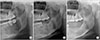
A 19-year-old female was referred to the dental hospital for further evaluation of a radiolucent lesion on the mandible. The patient was a non-smoker and had no underlying disease. A multi-ocular lesion was observed in the left mandibular body at the time of the visit. The panoramic view showed multiple root resorption near the posterior teeth and an inferiorly displaced inferior alveolar nerve canal.(Fig. 3. A) On extra-oral examination, facial asymmetry with swelling involving the left side of the mandible was noted. Bone expansion occurred at the left mandibular body and the inferior border of the mandible. An incisional biopsy was performed under local anesthesia, and the tissue was sent for histopathological examination, which resulted in a diagnosis of ameloblastoma.
Surgical enucleation or segmental mandibulectomy was regarded as inappropriate considering the patient's age and the size of the lesion. Extraction of #37 and #38 was performed under local anesthesia. Decompression was performed for nine months before surgery. A decreased size of the lesion was observed compared to the size at the initial visit.(Fig. 3. B)
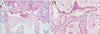
At the time of the operation, extraction of #35 and #36 and enucleation as a conservative surgery were performed, and removal of about 1 mm of bone near the lesion was also carried out. An excisional biopsy was performed, and the result revealed suggested unicystic ameloblastoma, mural type.(Fig. 4) One year after surgery, successful bone formation without any recurrence was observed.(Fig. 3. C)
3. Case 3
A 63-year-old male was referred from a local dental clinic for evaluation of swelling on the left side of his face. He was currently taking hypertension medicine. At the time of the patient's visit, edema was observed on the left side of his face and was accompanied by swelling on the left anterior ramus. Panoramic x-ray showed an expansive radiolucent lesion on the left mandibular ramus.(Fig. 5. A) Aspiration produced approximately 60 mL of straw-colored clear fluid. To reduce the size of the lesion, decompression was performed. Six months after decompression, the size of the lesion had decreased, and an incisional biopsy on the left retromolar region was carried out. The tissue examination results revealed an ameloblastoma. Decompression was constantly performed for 1 year; in a panoramic x-ray to observe the patient's progress, it was confirmed that the size had been reduced.(Fig. 5. B) One year after decompression, excision for ameloblastoma was performed under general anesthesia to remove tissue around the inferior alveolar nerve and connected to the mylohyoid muscle. An excisional biopsy was also performed, and the results were suggestive of unicystic ameloblastoma, follicular type.(Fig. 6)
One month postoperatively, the site of surgery was healing well, and numbness was resolving.(Fig. 5. C) Four months after the operation, panoramic views indicated marked new bone formation without any sign of recurrence at the operation site.
III. Discussion
None of the three patients relapsed into ameloblastoma during the follow-up period. The incisional biopsy performed under local anesthesia for one of the patients revealed a benign cyst. Based on this result, cyst enucleation and iliac bone graft were simultaneously performed under general anesthesia. Although the final results of the biopsy indicated unicystic ameloblastoma, mural type, the grafted bone settled without complication and showed a low chance of relapse. While the treatment of ameloblastoma was not a typical one, this case showed that a conservative surgical approach was an appropriate treatment.
Radical surgery was also not required for the female patient who developed ameloblastoma on the left mandible in light of her young age. However, to reduce the size, eight months of decompression were required. A conservative approach was therefore effective. We confirmed a reduction in the lesion's size on a radiograph, and a conservative surgical approach was carried out under general anesthesia. This patient also did not relapse.
The recommended treatment for this subtype of ameloblastoma is still controversial because treatment methods are dependent on the subtype. The choice of treatment is based not only on the apparent microscopic pattern on biopsy, but also on the location, size of the lesion, and age of the patient. Solid and multicystic ameloblastoma can be treated with surgery. Due to its ability to infiltrate the bone, it is regarded as a locally aggressive tumor67. Conservative resection, such as curettage and enucleation, is associated with a high recurrence rate (solid and multicystic, 60%–80%; unicystic, 30%–60%)8.
In contrast, unicystic ameloblastoma is thought to be less aggressive than solid or multicystic ameloblastoma. In most unicystic ameloblastoma cases, clinical and radiographic findings suggest that the lesion is an odontogenic cyst. These tumors are usually treated as cysts by enucleation.
According to Marx and Stern9, ameloblastoma in situ that develops in and is limited to the epithelial lining of a cyst and also microinvasive ameloblastoma that arises from the epithelial lining and proliferates into the connective tissue layer of the cyst should be treated with enucleation. However, the invasive type of ameloblastoma should be treated with resection in case the tumor has penetrated through the connective tissue of the cyst.
If the ameloblastic elements are confined to the lumen of the cyst either with or without intraluminal tumor extension, then cyst enucleation may be an adequate treatment option. The patient, however, should receive long-term follow-up treatment7. In the literature, the recurrence of unicystic ameloblastoma after conservative treatment is reported to be between 10% and 25%1011.
Because the rate of relapse is different for each treatment depending on the type of ameloblastoma, a clinical and radiological diagnosis must be made, and a biopsy is also necessary. However, as confirmed by numerous dissertations, since the relapse rate of unicystic ameloblastoma is relatively low compared to other types, conservative surgical treatment is the preferred method to avoid expansive losses that may occur with aggressive surgical treatment.

 XML Download