Journal List > J Adv Prosthodont > v.10(5) > 1105406

Jeon, Hwang, Kim, and Kim: Trueness and precision of scanning abutment impressions and stone models according to dental CAD/CAM evaluation standards

Abstract

PURPOSE

The purpose of the present study was to compare scanning trueness and precision between an abutment impression and a stone model according to dental computer-aided design/computer-aided manufacturing (CAD/CAM) evaluation standards.

MATERIALS AND METHODS

To evaluate trueness, the abutment impression and stone model were scanned to obtain the first 3-dimensional (3-D) stereolithography (STL) file. Next, the abutment impression or stone model was removed from the scanner and re-fixed on the table; scanning was then repeated so that 11 files were obtained for each scan type. To evaluate precision, the abutment impression or stone model was scanned to obtain the first 3-D STL file. Without moving it, scanning was performed 10 more times, so that 11 files were obtained for each scan type. By superimposing the first scanned STL file onto the other STL files one by one, 10 color-difference maps and reports were obtained; i.e., 10 experimental scans per type. The independent t-test was used to compare root mean square (RMS) data between the groups (α=.05).

RESULTS

The RMS±SD values of scanning trueness of the abutment impression and stone model were 22.4±4.4 and 17.4±3.5 µm, respectively (P<.012). The RMS±SD values of scanning precision of the abutment impression and stone model were 16.4±2.9 and 14.6±1.6 µm, respectively (P=.108).

CONCLUSION

There was a significant difference in scanning trueness between the abutment impression and stone model, as evaluated according to dental CAD/CAM standards. However, all scans showed high trueness and precision.

INTRODUCTION

With the advent of digital dentistry, it has become important to accurately evaluate dental computer-aided design/computer-aided manufacturing (CAD/CAM) devices.12 In this regard, it is vital that clinicians verify the accuracy of their scanners when using abutment stone models, and several investigations have focused on this topic.345 However, in patients with different tooth surface conditions, shapes, sizes, etc., it is almost impossible to find the same abutment. For this reason, it is difficult to evaluate scanning accuracy in such patients.67891011
Thus, researchers must evaluate scanning accuracy in cases that use the abutment stone model, which is standardized in dental prosthesis manufacturing. According to ISO 12836, a 3-unit bridge model has been used to evaluate the accuracy of dental CAD/CAM devices.12 However, few investigations have evaluated scanning accuracy using the abutment stone model, which is the most important prosthesis standard used for dental CAD/CAM systems.13
Conversely, many studies have evaluated the accuracy of abutment impression scanning, which has been used in recent digital dental prosthesis manufacturing.14 After the CAD file has been transmitted by the CAM milling machine to a 3D printer for manufacture of the dental prosthesis, the data obtained through direct impression scanning are saved in the CAD file.151617 However, to evaluate the accuracy of scanning of the abutment impression and stone model, it is necessary to verify both trueness and precision. Specifically, to evaluate trueness, one specimen should be scanned by a single scanner; after the initial scanning, the specimen is removed completely from the scanner table, re-fixed to the table again, and rescanned. The scan data obtained by repeating this process several times can be superimposed and verified. To assess precision, one specimen is scanned several times by a single scanner without being removed. The scan data obtained by repeating the process can be superimposed and verified, as with the above trueness verification method.1819
In general dentistry, the trueness and precision of abutment stone model scanning are higher than those of impression scanning.420212223 However, few investigations have evaluated scanning trueness and precision using dental CAD/CAM evaluation standards. This is an important concern in current digital CAD/CAM dentistry.
Therefore, the purpose of the present study was to compare trueness and precision between impression scanning and abutment stone model scanning according to dental CAD/CAM evaluation standards.

MATERIALS AND METHODS

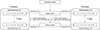
To prepare the abutment impression and stone model according to the dental CAD/CAM evaluation standards, the specimens were produced as shown in Fig. 1. Briefly, the abutment (Geomagic Design X 2016, 3D Systems, Cary, NC, USA) was designed with an upper-end diameter of 5.2 mm. The lower part had a diameter of 8 mm, a crown length of 10 mm, and a crown inclination of 8°.12 The milling process used the designed CAD data, and a titanium abutment model was produced. Using an extra light body (Aquasil Ultra, Dentsply, York, PA, USA), which has the best fluidity and refinement among silicone rubber impression materials, a duplicate impression of the titanium abutment model could be obtained.
The abutment stone model was then created using the duplicated impression. The model was made using gypsum (Snow Rock 3D Scan Stone, DK Mungyo, Gimhae, Korea), which is an optimal material for scanning.
To evaluate the trueness and precision of abutment impression and stone model scanning, a blue LED scanner was used (Identica blue, Medit, Seoul, Korea). This is a recently developed scanner, and it reportedly has a smaller scan error and higher scanning accuracy than conventional scanners.672425 Comparative evaluation of the trueness and precision of the abutment impression scanning were then compared with those of the stone model scanning according to dental CAD/CAM evaluation standards. To evaluate the trueness of abutment impression scanning, the impression was fixed on the scanner table. It was then scanned to obtain the first 3-dimensional (3-D) stereolithography (STL) file, named TI_1. Next, the impression was removed and refixed to the table and scanned again. This operation was repeated 10 times to obtain 10 more STL files (TI_2-11), and a total of 11 STL files were obtained (TI_1-11). To evaluate the trueness of abutment stone model scanning, the same operations were performed to obtain 11 STL files (TS_1-11).
In contrast, to evaluate the precision of abutment impression scanning, the impression was fixed to the scanner table and scanned to obtain the first 3-D STL file (PI_1). It was then scanned 10 more times without being moved, and 10 more STL files were obtained (PI_2-11), resulting in a total of 11 STL files (PI_1-11). To evaluate the precision of abutment stone model scanning, the same operations were performed to obtain files (PS_1-11).
In all STL files, unnecessary and inaccurate parts were deleted.26272829 To verify the trueness and precision of the 3-D STL files, the following method was employed. First, the trueness of the abutment impression scanning was evaluated using 3-D superimposing software (Geomagic Verify 2015, 3D Systems, Cary, NC, USA). By superimposing the first scanned STL file (TI_1; control scan) onto the other STL files (TI_2-11; experimental scans) one by one, 10 color-difference maps and reports were obtained. To evaluate the trueness of the abutment stone model scanning, the same operations were performed to obtain 10 color-difference maps and reports.
Similarly, to evaluate the precision of the abutment impression scanning, the first scanned STL file (SI_1; control scan) was superimposed onto the other STL files (SI_2-11; experimental scans) one by one to obtain 10 color-difference maps and reports. To evaluate the precision of the abutment stone model scanning, the same operations were performed to obtain 10 color-difference maps and reports.
In this way, in the report obtained using 3-D superimposing software, 10 quantitative root mean square (RMS) values were obtained for each abutment impression and stone model using the equation below:
RMS=m=1nx1,mx2,m2njap-10-335-e001
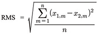
That is, when two scans were superimposed, the square of the phase difference between a number of points in 3-D space was calculated (x-, y-, and z-axis). The sum of these squares was divided by the number of points, and RMS was calculated as the square root of this value. This may be a more reliable and accurate value than a general arithmetic mean because the difference between each data point is represented by both a positive value (red in the color-difference map; Fig. 2) and a negative value (blue in the color-difference-map; Fig. 2). The reliability of arithmetic means is limited in cases of simple sums.130
With regards to statistical analysis, an independent t-test was used to verify the significance of the differences between the groups. IBM SPSS version 22.0 for Windows (IBM SPSS Inc., Chicago, IL, USA) was used. The level of significance of α error was 0.05.

RESULTS

Table 1 shows the RMS (± SD) values of the trueness and precision comparisons between the abutment impression and stone model, which were carried out according to dental CAD/CAM evaluation standards. There was a significant difference in terms of trueness between the abutment impression and stone model scanning (P < .012), but not in terms of precision (P = .108).
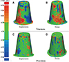
Fig. 2 shows the color-difference map comparing trueness and precision between the abutment impression and stone model according to dental CAD/CAM evaluation standards.
The color-difference map of trueness showed many positive (red) errors and negative (blue) errors throughout both the abutment impression (Fig. 2A) and the axis part of the stone model (Fig. 2B).
The color-difference map of precision showed some positive (red) errors and negative (blue) errors in the axis parts of both the abutment impression (Fig. 2C) and stone model (Fig. 2D).

DISCUSSION

In the present study, we began with the null hypothesis that trueness and precision do not differ between the abutment impression and stone model, as measured using dental CAD/CAM evaluation standards.
There was a significant difference in trueness between the abutment impression and stone model (P < .012) perhaps due to a high probability of error at the time of scanning, as some shadows were generated when the impression was scanned. Despite this significant difference, few errors occurred, and each was only of about 5 µm in size.
On the other hand, there was no significant difference in scanning precision between the abutment impression and stone model (P = .108). In general, it has been taken for granted in dental CAD/CAM that the abutment stone model is superior to the abutment impression in terms of scanning accuracy.218 However, we found no significant difference, and only a few errors occurred, each of which was only about 2 µm in size.
The color-difference map of trueness showed many positive (red) errors and negative (blue) errors in the axis parts of both the abutment impression and stone model (Fig. 2A, Fig. 2B). This is because the area into which the LEDs are projected at the time of scanning expands in the direction of the major axis, resulting in a high probability of scan error.731
The color-difference map of precision showed some positive (red) errors and negative (blue) errors in the axis parts of both the abutment impression and stone model (Fig. 2C, Fig. 2D). This is because the blue light scanner used in this study uses short-wavelength light, so it is hardly affected by the factors that cause scanning errors, such as the shape, color, and size of the scanning object.72224
To obtain more reliable results, we made some additional effort in experimental design in the present study. First, the stone model was produced using 3-D scan stone, which is an optimal material for dental scanners, as was shown recently. In addition, we evaluated the trueness and precision of abutment impression and stone model scanning using 3-D superimposition software, which has been used not only in dentistry, but also in engineering, medicine, pharmacy, and other fields. In fact, the software is recognized worldwide for its reliability.32333435
Nonetheless, there were some limitations to this research. First, we failed to adequately explain errors due to reflection, refraction, and scattering of light while using the blue light scanner, which is only one type of optical scanner.253136 Next, trueness and precision were evaluated using the 3-D superimposing method rather than the conventional 2-D measurement method. However, we could not explain the best fit alignment process used to minimize and verify the errors between the data2437 because it is difficult to judge abutment impression and stone model scanning, or the best fit alignment process, in terms of errors in qualitative and quantitative data obtained using 3-D superimposition software.223839
Therefore, future research must seek to reduce errors in trueness and precision evaluation using 3-D superimposing software, and continuing efforts must be made to improve scanning quality using abutment impressions and stone models, in accordance with dental CAD/CAM evaluation standards.

CONCLUSION

There was a significant difference in scanning trueness between the abutment impression and stone model, as evaluated according to dental CAD/CAM standards. However, all scans showed high trueness and precision. The results of this research will be useful in digital CAD/CAM dentistry.

Figures and Tables

Fig. 1

Experimental schematic diagram of this study.

jap-10-335-g001
Fig. 2

Qualitative comparison data, represented using a 3-dimensional color map of both trueness and precision (impression vs. stone model).

jap-10-335-g002
Table 1

Quantitative comparison of trueness and precision of scanning impression and stone model (10 images per scan type)

jap-10-335-i001
Impression Stone P value
Trueness (RMS ± SD) 22.4 ± 4.4 17.4 ± 3.5 < .012
Precision (RMS ± SD) 16.4 ± 2.9 14.6 ± 1.6 .108

Unit: µm, RMS: root mean square, SD: standard deviation

Notes

This study was supported by grants from Korea University and Kyung-Dong University. The authors appreciate the representatives and staff of Medit Inc. for providing access to their blue LED scanner.

References

1. Jeong ID, Kim WC, Park J, Kim CM, Kim JH. Ceramic molar crown reproducibility by digital workflow manufacturing: An in vitro study. J Adv Prosthodont. 2017; 9:252–256.
crossref
2. Luthardt RG, Loos R, Quaas S. Accuracy of intraoral data acquisition in comparison to the conventional impression. Int J Comput Dent. 2005; 8:283–294.
3. Carbajal Mejía JB, Wakabayashi K, Nakamura T, Yatani H. Influence of abutment tooth geometry on the accuracy of conventional and digital methods of obtaining dental impressions. J Prosthet Dent. 2017; 118:392–399.
4. González de Villaumbrosia P, Martínez-Rus F, García-Orejas A, Salido MP, Pradíes G. In vitro comparison of the accuracy (trueness and precision) of six extraoral dental scanners with different scanning technologies. J Prosthet Dent. 2016; 116:543–550.
5. Jeon JH, Kim HY, Kim JH, Kim WC. Accuracy of 3D white light scanning of abutment teeth impressions: evaluation of trueness and precision. J Adv Prosthodont. 2014; 6:468–473.
crossref
6. Renne W, Ludlow M, Fryml J, Schurch Z, Mennito A, Kessler R, Lauer A. Evaluation of the accuracy of 7 digital scanners: An in vitro analysis based on 3-dimensional comparisons. J Prosthet Dent. 2017; 118:36–42.
7. Lee JJ, Jeong ID, Park JY, Jeon JH, Kim JH, Kim WC. Accuracy of single-abutment digital cast obtained using intraoral and cast scanners. J Prosthet Dent. 2017; 117:253–259.
crossref
8. Bramanti E, Cervino G, Lauritano F, Fiorillo L, D'Amico C, Sambataro S, Denaro D, Famà F, Ierardo G, Polimeni A, Cicciù M. FEM and von Mises analysis on prosthetic crowns structural elements: Evaluation of different applied materials. ScientificWorldJournal. 2017; 2017:1029574.
crossref
9. Cicciù M, Cervino G, Bramanti E, Lauritano F, Lo Gudice G, Scappaticci L, Rapparini A, Guglielmino E, Risitano G. FEM analysis of mandibular prosthetic overdenture supported by dental implants: evaluation of different retention methods. Comput Math Methods Med. 2015; 2015:943839.
crossref
10. Cicciù M, Bramanti E, Cecchetti F, Scappaticci L, Guglielmino E, Risitano G. FEM and Von Mises analyses of different dental implant shapes for masticatory loading distribution. Oral Implantol (Rome). 2014; 7:1–10.
11. Cicciú M, Bramanti E, Matacena G, Guglielmino E, Risitano G. FEM evaluation of cemented-retained versus screw-retained dental implant single-tooth crown prosthesis. Int J Clin Exp Med. 2014; 7:817–825.
12. ISO 12836. Dentistry - Digitizing devices for CAD/CAM systems for indirect dental restorations - Test methods for assessing accuracy. Geneva, Switzerland: International Standards for Organization (ISO);2015. Accessed March 2, 2016. Available from: http://www.iso.org/iso/store.html.
13. Jeong ID, Lee JJ, Jeon JH, Kim JH, Kim HY, Kim WC. Accuracy of complete-arch model using an intraoral video scanner: An in vitro study. J Prosthet Dent. 2016; 115:755–759.
14. Jeon JH, Lee KT, Kim HY, Kim JH, Kim WC. White light scanner-based repeatability of 3-dimensional digitizing of silicon rubber abutment teeth impressions. J Adv Prosthodont. 2013; 5:452–456.
crossref
15. Kim DY, Lee HN, Kim JH, Kim HY, Kim WC. Evaluation of marginal and internal gaps in single and three-unit metal frameworks made by micro-stereolithography. J Adv Prosthodont. 2017; 9:239–243.
crossref
16. Lee WS, Lee DH, Lee KB. Evaluation of internal fit of interim crown fabricated with CAD/CAM milling and 3D printing system. J Adv Prosthodont. 2017; 9:265–270.
crossref
17. Kournetas N, Spintzyk S, Schweizer E, Sawada T, Said F, Schmid P, Geis-Gerstorfer J, Eliades G, Rupp F. Comparative evaluation of topographical data of dental implant surfaces applying optical interferometry and scanning electron microscopy. Dent Mater. 2017; 33:e317–e327.
crossref
18. Nedelcu RG, Persson AS. Scanning accuracy and precision in 4 intraoral scanners: an in vitro comparison based on 3-dimensional analysis. J Prosthet Dent. 2014; 112:1461–1471.
19. ISO-5725-1. Accuracy (trueness and precision) of measurement methods and results. Part 1: General principles and definitions. Geneva, Switzerland: International Standards for Organization (ISO);1994. Accessed December 22, 2015. Available at: http://www.iso.org/iso/store.html.
20. Ender A, Mehl A. Accuracy of complete-arch dental impressions: a new method of measuring trueness and precision. J Prosthet Dent. 2013; 109:121–128.
crossref
21. Lauritano F, Runci M, Cervino G, Fiorillo L, Bramanti E, Cicciù M. Three-dimensional evaluation of different prosthesis retention systems using finite element analysis and the Von Mises stress test. Minerva Stomatol. 2016; 65:353–367.
22. Jeon JH, Jung ID, Kim JH, Kim HY, Kim WC. Three-dimensional evaluation of the repeatability of scans of stone models and impressions using a blue LED scanner. Dent Mater J. 2015; 34:686–691.
crossref
23. Cicciù M, Risitano G, Maiorana C, Franceschini G. Parametric analysis of the strength in the “Toronto” osseous-prosthesis system. Minerva Stomatol. 2009; 58:9–23.
24. Jeon JH, Kim DY, Lee JJ, Kim JH, Kim WC. Repeatability and reproducibility of individual abutment impression, assessed with a blue light scanner. J Adv Prosthodont. 2016; 8:214–218.
crossref
25. Kim DY, Jeon JH, Kim JH, Kim HY, Kim WC. Reproducibility of different arrangement of resin copings by dental microstereolithography: Evaluating the marginal discrepancy of resin copings. J Prosthet Dent. 2017; 117:260–265.
crossref
26. Ausiello P, Ciaramella S, Fabianelli A, Gloria A, Martorelli M, Lanzotti A, Watts DC. Mechanical behavior of bulk direct composite versus block composite and lithium disilicate indirect Class II restorations by CAD-FEM modeling. Dent Mater. 2017; 33:690–701.
crossref
27. Ausiello P, Ciaramella S, Garcia-Godoy F, Gloria A, Lanzotti A, Maietta S, Martorelli M. The effects of cavity-margin-angles and bolus stiffness on the mechanical behavior of indirect resin composite class II restorations. Dent Mater. 2017; 33:e39–e47.
crossref
28. Li J, Yuan P, Chang CM, Ho DC, Lo YF, Shen S, Kim D, Teichgraeber JF, Alfi DM, Gateno J, Xia JJ. New approach to establish an object reference frame for dental arch in computer-aided surgical simulation. Int J Oral Maxillofac Surg. 2017; 46:1193–1200.
crossref
29. Lebon N, Tapie L, Duret F, Attal JP. Understanding dental CAD/CAM for restorations - dental milling machines from a mechanical engineering viewpoint. Part A: chairside milling machines. Int J Comput Dent. 2016; 19:45–62.
30. Kim CM, Jeon JH, Kim JH, Kim HY, Kim WC. Three-dimensional evaluation of the reproducibility of presintered zirconia single copings fabricated with the subtractive method. J Prosthet Dent. 2016; 116:237–241.
crossref
31. Jeon JH, Choi BY, Kim CM, Kim JH, Kim HY, Kim WC. Three-dimensional evaluation of the repeatability of scanned conventional impressions of prepared teeth generated with white- and blue-light scanners. J Prosthet Dent. 2015; 114:549–553.
crossref
32. Vecsei B, Joós-Kovács G, Borbély J, Hermann P. Comparison of the accuracy of direct and indirect three-dimensional digitizing processes for CAD/CAM systems - An in vitro study. J Prosthodont Res. 2017; 61:177–184.
crossref
33. Moser M, Schmid R, Schindel R, Hildebrandt G. Patient-specific polymethylmethacrylate prostheses for secondary reconstruction of large calvarial defects: A retrospective feasibility study of a new intraoperative moulding device for cranioplasty. J Craniomaxillofac Surg. 2017; 45:295–303.
crossref
34. Kim DS, Lee B, Banks SA, Hong K, Jang YH. Comparison of dynamics in 3D glenohumeral position between primary dislocated shoulders and contralateral healthy shoulders. J Orthop. 2017; 14:195–200.
crossref
35. Liu L, Li H, Cui Y, Li R, Meng F, Ye Z, Zhang X. Calcium channel opening rather than the release of ATP causes the apoptosis of osteoblasts induced by overloaded mechanical stimulation. Cell Physiol Biochem. 2017; 42:441–454.
crossref
36. Martorelli M, Ausiello P, Morrone R. A new method to assess the accuracy of a Cone Beam Computed Tomography scanner by using a non-contact reverse engineering technique. J Dent. 2014; 42:460–465.
crossref
37. Flügge TV, Schlager S, Nelson K, Nahles S, Metzger MC. Precision of intraoral digital dental impressions with iTero and extraoral digitization with the iTero and a model scanner. Am J Orthod Dentofacial Orthop. 2013; 144:471–478.
crossref
38. Persson AS, Andersson M, Odén A, Sandborgh-Englund G. Computer aided analysis of digitized dental stone replicas by dental CAD/CAM technology. Dent Mater. 2008; 24:1123–1130.
crossref
39. Quaas S, Rudolph H, Luthardt RG. Direct mechanical data acquisition of dental impressions for the manufacturing of CAD/CAM restorations. J Dent. 2007; 35:903–908.
crossref
TOOLS
ORCID iDs

Jin-Hun Jeon
https://orcid.org/0000-0003-2833-8946

Seong-Sig Hwang
https://orcid.org/0000-0003-1716-2594

Ji-Hwan Kim
https://orcid.org/0000-0003-3889-2289

Woong-Chul Kim
https://orcid.org/0000-0002-6730-4960

Similar articles