

 PDF
PDF ePub
ePub Citation
 Print
Print


INTRODUCTION
Gallbladder adenomyomatosis is a relatively common disease, characterized by proliferation of Rokitansky-Aschoff sinuses within the hypertrophied muscularis. Rokitansky-Aschoff sinus is a mucosal outpouching structure that sits within the muscular layer of gallbladder. The proliferation and hypertrophy of these structures cause wall thickening of the gallbladder.
Localized type of adenomyomatosis rarely presents as a polypoid lesion, and sometimes mimics an advanced gallbladder cancer (12). Herein, we describe an unusual manifestation of gallbladder adenomyomatosis showing polypoid growth through the colon, resulting in preoperatively misdiagnosed as an advanced hepatic flexure colon cancer.
CASE REPORT
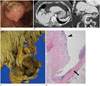
A 62-year-old woman was referred to our hospital for an incidentally found colonic polyp on a screening colonoscopy. She had no relevant past history and was symptomless. Physical examination was unremarkable. Laboratory tests were within the normal limits, except for mildly elevated C-reactive protein (0.91 mg/dL). A repeat colonoscopy for a planned polypectomy revealed a polypoid mass in the hepatic flexure of the colon (Fig. 1A). The mucosal surface of the mass showed different nature from that of the normal colonic mucosa, and had an ulceration as well as denudation at the stalk and the base of the polyp. Under the suspicion for a malignant polyp, indigo carmine solution was injected in the submucosal layer of the nearby colon for polypectomy. Since the polyp failed to be lifted after the injection, the patient was further consulted for the surgical resection with the diagnostic impression of a colon cancer that had invaded beyond the submucosal layer. The biopsy of the polyp obtained during the second colonoscopy came back as an inflammatory polyp.
A contrast-enhanced computed tomography (CT) scan was performed for further evaluation. Abdominal CT depicted a 3 cm heterogeneously enhancing polypoid intraluminal mass in the hepatic flexure of the colon. The mass was extending to the adjacent gallbladder fundus accompanying a 1 cm gallbladder stone. The polyp had lobulated surface, without an overlying colonic mucosa. The mass was closely abutting the liver causing tethered appearance of the liver surface (Fig. 1B). There were no significantly enlarged regional lymph nodes. Based on the CT findings, colon cancer with gallbladder and liver invasion was strongly suggested.
The patient underwent right hemicolectomy. In line with the CT images, the colonic mass was found to be adjoining the gallbladder during the surgery. As the mass was adherent to the liver parenchyma, radical cholecystectomy which removes both the gallbladder and a part of the liver was performed.
Gross pathologic specimen revealed a polypoid mass in the colonic lumen that had protruded from the gallbladder fundus (Fig. 1C). Histopathologically, the mass was composed of polypoid growth of gallbladder adenomyomatous hyperplasia. The surface of the mass was covered with a gallbladder mucosa and was continuous with colonic mucosa (Fig. 1D). There were scanty inflammatory cells and fibrosis around the protruded gallbladder adenomyomatosis and adjacent colonic wall. No malignant cell was detected.
DISCUSSION
Adenomyomatosis or adenomyomatous hyperplasia of the gallbladder is a common benign disease, identified in approximately 9% of specimens obtained after cholecystectomy (3). Gallbladder adenomyomatosis is divided into three types depending on the location or the extent of involvement: diffuse, segmental, and localized. The most common type is localized or focal adenomyomatosis presenting as a crescentic wall thickening of the gallbladder, usually at the fundus. Localized form of the adenomyomatous hyperplasia is rarely polypoid, and sometimes this polypoid appearance is mistaken for an advanced gallbladder cancer (12). However, there has been no report of gallbladder adenomyomatosis mimicking colon cancer, to our knowledge.
On contrast-enhanced CT scan, a polypoid soft tissue mass was extending from the colonic lumen to the adjacent gallbladder and liver in our case. Because the colon is the second most commonly invaded organ in gallbladder cancer (15%) and, though less common, a gallbladder invasion is also observed in the colon cancer (2.7%), either colon or gallbladder cancer should be the first differential diagnosis (45). Radiologic finding of an advanced gallbladder cancer with colonic invasion and vice versa is a soft tissue mass and/or wall thickening continuously involving both the colon and the gallbladder. It may be difficult to identify the primary site of the cancer in an advanced disease due to overlapping imaging features. However, scrutinizing the epicenter of the mass and confirming the intact colonic mucosa on colonoscopy may help in identifying the primary cancer.
In our case, most part of the lesion had protruded into the colonic lumen and was covered with a non-colonic mucosa to resemble a hepatic flexure colon cancer. One possible mechanism for a benign gallbladder lesion to penetrate into the colon is a cholecystocolic fistula, although there was neither definite fistulous tract nor extensive fibrosis that may suggest fibrotic obliteration of the fistula in the gross and microscopic evaluations in our case. Nevertheless, we presume cholecystocolic fistula as a feasible mechanism since the adenomyomatosis covered with gallbladder mucosa had protruded into the colonic lumen, and the gallbladder mucosa was clearly demarcated from the adjacent colonic mucosa. Thus, though not sure, a preceding cholecystocolic fistula may have facilitated extension of the gallbladder adenomyomatosis through it and later could have been obliterated.
A cholecystocolic fistula is a rare complication observed in 0.06–0.14% of patients with biliary disease (67). Various benign and malignant gallbladder diseases can form cholecystocolic fistula (8), and even mimic a colon cancer in case of a gallbladder cancer that had invaded the colon (9). The pathogenesis of fistula formation in adenomyomatosis is unclear. As Rokitansky-Aschoff sinus is considered as an analogous to diverticulum of the colon, increased intraluminal pressure of the gallbladder can induce perforation of a Rokitansky-Aschoff sinus. This may result in a localized abscess formation and consecutive fistulation, particularly in condition with gallstones or associated chronic gallbladder inflammation (10). Our patient also had a gallstone, and it may have elevated intraluminal pressure leading to a repetitive inflammation. One interesting finding was that the mass had a gallbladder mucosa on the luminal side of the colon. Though uncertain, adenomyomatous hyperplasia of smooth muscle layer could have pushed the gallbladder mucosa through the cholecystocolic fistula to the colonic side.
In conclusion, we present a unique case of polypoid gallbladder adenomyomatosis mimicking a hepatic flexure colon cancer owing to its bulginess and non-colonic mucosal surface. Physicians and radiologists should be aware that benign gallbladder disease can mimic a mucosal origin colon cancer without cholecystocolic fistula and be cautious when managing hepatic flexure colonic polyps especially in patients with chronic gallbladder inflammation.
 XML Download