

 PDF
PDF ePub
ePub Citation
 Print
Print


INTRODUCTION
Toxocariasis is a zoonotic infestation from parasite Toxocara canis (T. canis) and cati (T. cati) that manifests in variable parts of human body including the liver, lungs, eyes, heart, and the brain. The specific form of toxocariasis involving the systemic organs such as the liver, lungs and the gastrointestinal tract is named visceral larva migrans (VLM) and it presents clinical symptoms such as abdominal pain, fever, cough and wheezing (1). VLM mainly manifests as infiltrations in the liver (2) or as gastroenteritis (3) and ascites (4) in the abdomen, but rarely as generalized lymphadenopathy (15). There have been rare reports of toxocariasis presented as multiple lymphadenopathies in the chest (1) or neck areas (5) but not in the abdomen. Therefore, we report a case of toxocariasis presented as multiple conglomerated lymphadenopathy that was initially misinterpreted as lymphoma on abdomen computed tomography (CT).
CASE REPORT
A 43-year-old female was referred to our hospital for abnormal findings on outside abdomen and pelvis CT scan. The patient had no special past medical or family history and the general physical examination was unremarkable. A thorough history taking of the patient revealed that the patient had a career as a pet groomer at an animal hospital and raised a cat and a stray dog at home.
The patient's white blood cell count was 3.51 × 10³/uL with 6.4% neutrophils, 5.3% lymphocytes, and 84.8% eosinophils (normal range: 0–5%). The peripheral blood smear showed eosinophilia of 83%, absolute number of 2.84 × 10⁹/L (normal absolute number: < 0.5 × 10⁹/L). Lactate dehydrogenase level was 744 IU/L (normal range: 0–480) and serum IgE level was 154 IU/mL (normal range: < 100 IU/mL). Erythrocyte sedimentation rate was 25 mm/hr and C-reactive protein was 3.21 mg/L, both within normal limit. The lab results of electrolytes, liver function, renal function and urine analysis were within normal limit. Since the patient had a career involving multiple contacts with animals, allergy test was carried out measuring IgE levels for possible allergens of animals and pollen. The allergy tests were within normal range. Also, helminthic serologic tests were carried out using enzyme-linked immunosorbent assay (ELISA) kit to detect specific IgG antibodies for specific parasites. T. canis specific IgG titer was positive with increased value of 1.364 (cutoff value 1.027) whereas other parasite infections such as Cysticercus, Sparganum, Paragonimus westermani, and Clonorchis sinensis were excluded.
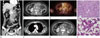
Abdominal CT revealed multiple conglomerated enlarged lymph nodes in gastro hepatic, portocaval, aortocaval, paraaortic, mesenteric, and bilateral inguinal areas showing homogenous enhancement (Fig. 1A). The maximal diameter of the conglomerated, enlarged mesenteric lymph nodes was larger than 10 cm in axial view and larger than 15 cm in coronal view. The enlarged lymph nodes showed the ‘sandwich sign’ by surrounding mesenteric vessels (Fig. 1B). The patient did not accompany organomegaly, bowel wall thickening or ascites. The chest CT scan revealed enlarged bilateral cervical, hilar, mediastinal and axillary lymph nodes with similar features (Fig. 1C). The initial radiologic impression for both the abdomen and chest CT suggested lymphoma.
The patient underwent positron emission tomography (PET)-CT scan with fluorine-18-fluorodeoxyglucose and found multiple hyper metabolic lymph nodes in the neck, axilla and the abdomen (Fig. 1D). The PET-CT report suspected lymphoma involvements in the affected lymph nodes as well, consistent with the abdomen CT report. Furthermore, the patient went through axillary lymph node and bone marrow biopsy, and reactive eosinophilic hyperplasia was pathologically diagnosed (Fig. 1F). Conclusively, the patient was diagnosed as toxocariasis by excluding multiple probable causes for eosinophilia and detecting specific IgG to T. canis.
The patient was treated with antihelminthic drugs, praziquantel (600 mg, three times a day- for two days and albendazole (400 mg, twice a day) for two weeks, followed with oral steroids (30 mg, twice a day) for one week. Progressive improvement of eosinophilia was observed from initial count of 84.8% to 6.3% at follow up and the lactate dehydrogenase level was normalized to 378 IU/L. At follow up CT scan taken four months later, the lymph node enlargements had dramatically decreased in size (Fig. 1E).
DISCUSSION
Toxocariasis is a parasitic zoonosis from roundworm infections of T. canis and T. cati which live in a dog's small intestine. Human are infected by ingesting ova from contaminated soil or larvae from uncooked meat (cow, pig, lamb, and chickens). The ingested ova or larva invades the small bowel and travels through the portal venous system to reach the liver and spread to the lungs, eyes and the brain (6).
There are two main manifestations of toxocariasis: VLM and ocular larva migrans. VLM is the systemic exhibition showing common clinical features of peripheral eosinophilia, abdominal pain, hepatosplenomegaly, fever, and hypergammaglobulinemia (67).
Toxocariasis is diagnosed definitively by visually confirming the larva in infected tissues, but this method has limitations of being insensitive and time-consuming. Therefore the most commonly used indirect diagnostic method for toxocariasis is currently ELISA test for detecting anti-Toxocara IgG antibodies (89). After diagnosis, patients are treated with antiparasitic drugs often in combination with corticosteroids to suppress allergic manifestations (10). Our case was diagnosed as toxocariasis with the ELISA method showing increased titers for Toxocara specific IgG. Consequently, the patient was treated with antiparasitic drugs in combination with steroids.
Radiologic findings of VLM overlaps with those caused from eosinophilia, showing eosinophilic infiltrations across variable visceral organs. However, VLM predominantly involves the liver and occasionally involves the gastrointestinal tract or the lungs, as it spreads via the portal system. The affected areas are presented as hepatic eosinophilic infiltrations (2) or eosinophilic gastroenteritis with ascites (34). There have been two case reports on the rare manifestation of toxocariasis as generalized lymphadenopathies (15). Szczepański et al. (5) reported on lymphadenopathies involving the cervical, supraclavicular and inguinal areas with sizes about 0.5–2 cm, but did not offer radiologic images. Bachmeyer et al. (1) reported bilateral hilar and mediastinal lymphadenopathies displayed in the chest radiograph and CT scan with sizes about 2–5 cm in diameter, but the involved lymph nodes were limited to the chest. However, our case showed diffuse conglomerated lymphadenopathies involving multiple areas including the lower neck, bilateral axilla and the abdomen, which were first misinterpreted as lymphoma involvements.
It is important that the radiologists are aware of this rare form of toxocariasis- associated hypereosinophilia. Careful history taking and serologic testing for parasitic infestations are required for accurate diagnosis.
 XML Download