Abstract
The falciform ligament is a hepatic suspensory ligament that extends from the umbilicus to the diaphragm, containing the ligamentum teres and a vestigial remnant of the umbilical vein. Among the rarely-occurring pathologies of the falciform ligament, which include ligament cyst, tumor, abnormal vascularization, and congenital ligament defect, a falciform ligament abscess is even more sporadic. Accordingly, the definitive diagnosis of the falciform ligament abscess is rather challenging and may easily be misinterpreted as an infected choledochal cyst or a liver abscess. We present a 25-day-old infant with the falciform ligament abscess, which developed after the umbilical venous catheter insertion and was successfully treated with percutaneous drainage and antibiotic administration.
The peritoneal folds, known to include the ligaments, the mesentery and omenta, which encompass blood vessels and lymphatics, connect the intraperitoneal organs to the abdominal wall, and provide a pathway for the spread of disease (1). The falciform ligament is one of the suspensory ligaments of the liver, which contains the ligament teres and an umbilical vein remnant (2). Ligaments generally provide a potential space for the spread of disease, yet a primary falciform ligament-related disease is quite rare. A falciform ligament abscess is even rarer than any other falciform ligament-related disorders. There were few reports of the falciform ligament abscess after omphalitis: one extended from omphalitis through the paraumbilical venous network (3) and the others resulted from contiguous spread of the infection via the round ligament (4). In this report, we present a 25-day-old infant with the falciform ligament abscess that developed after umbilical venous catheter (UVC) insertion, which was effectively managed with percutaneous drainage and antibiotic treatment.
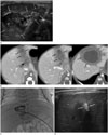
A 25-day-old boy, weighing 2320 g, was born to a gravida 1 para 1 mother at about 39 weeks of gestation by normal spontaneous vaginal delivery at a local hospital. The boy was stained with meconium (4+) at birth, and had difficulty breathing and decreased limb motions. The initial laboratory test showed a white blood cell (WBC) count of 9900/mm3 and a serum C-reactive protein (CRP) of 127.4 mg/L (< 5 mg/L). He was suspected of having birth asphyxia, and transferred to the neonatal intensive care unit. On hospital day 1, the boy had an UVC inserted in order to infuse total parenteral nutrition and antibiotics. On hospital day 6, his UVC was removed. There were no umbilical discharge, periumbilical erythema, or umbilical necrosis before and after UVC removal. The follow-up laboratory test taken on hospital day 13 revealed a WBC count of 11680/mm3. On hospital day 17, he developed a fever and abdominal distension as his leukocytosis was exacerbated, showing the WBC count as high as 22310/mm3. The transabdominal ultrasound (US) (Fig. 1A) and computed tomography (CT) (Fig. 1B) revealed a 3.7 × 4.2 cm abscess along the course of the falciform ligament.
Percutaneous catheter drainage (PCD) was performed to treat the falciform ligament abscess (Fig. 1C). US guided percutaneous abscess puncture was done with 18G needle (Trocar Needle, COOK Inc., Bloomington, IN, USA) in epigastric area after local anesthesia under sedative anesthesia. After sampling aspiration, abscess cavitography was taken with injection of 50% diluted contrast material (Bonorex 350 inj; CMS, Seoul, Korea) through the needle. Under fluoroscopy guidance, 0.035-inch angled guide wire (Radifocus, Terumo, Tokyo, Japan) was inserted through the needle and the needle was taken out. 8.5 Fr pig-tail catheter (Multipurpose Drainage Catheter, COOK Inc.) was inserted into the abscess cavity over the guide wire which was then pulled out. The catheter was fixed to the skin and total 13 mL of bloody pus was drained. The aspirated pus sample was sent to the laboratory for culture study and the isolated pathogen was Enterobacter cloacae.
This patient's WBC count was normalized and his serum CRP improved 10 days after the PCD procedure and the average daily drainage was 3 mL. The catheter was removed on day 9 and the follow-up transabdominal US carried out one month after the catheter removal showed complete regression of the falciform ligament (Fig. 1D).
The characteristic US appearance of the falciform ligament is an echogenic focus at the junction of the quadrate and left lobes of the liver. Recognition of the typical location of the falciform ligament is especially important to distinguish it from other intrahepatic masses (5).
The falciform ligament may be affected secondarily by an infection from surrounding organs such as the gallbladder and the liver (67). Moreover, the falciform ligament abscess may easily be misdiagnosed as an infected choledochal cyst or a liver abscess (8). In the current case, the abscess was shown in the area between the quadrate and left lobes of the liver, which is the typical location for the falciform ligament. The CT and US images showed no evidence of either cholecystitis or hepatitis. Furthermore, no intrahepatic and common bile duct dilatations were revealed on CT and US images. Therefore, based on these imagery findings, the possibility of an infected choledochal cyst, a liver mass or an infection spread from the gallbladder and the liver to the falciform ligament could be excluded.
The pathophysiologic mechanism of the falciform ligament abscess can be explained either by the contiguous spread of an infection via the round ligament (4), or by the extension through the paraumbilical venous network (3). In the current case, the patient did not show any direct signs and symptoms (i.e., umbilical discharge, erythema, necrosis, or umbilical cellulitis) of neonatal omphalitis as defined by Sawadekar (9). Moreover, the patient's systemic signs of an infection were distinctive and his leukocytosis got worse after the removal of the UVC. Thus, the prospect of having an infectious spread of the venous network is more likely than that of having a direct spread from the round ligament, in this case, with respect to the probable pathophysiologic mechanism of the falciform ligament abscess.
Virtually all case reports on the issue of the falciform ligament abscess indicate that excision of the abscess in the falciform ligament is the treatment of choice. By the same token, the authors of those reports insist that surgical excision should be considered for the initial treatment for the falciform ligament abscess (3478). Nonetheless, in the current case, the pediatrician in charge of this patient decided to treat the baby with PCD initially because the baby was too young to endure surgery and already afflicted with a hypoxic brain injury. Unlike the previous reports, his falciform ligament abscess vanished, regressing completely after PCD. The baby was free of signs and symptoms of infection. Therefore, PCD may be the good treatment option for the falciform ligament abscess in cases where surgery may not be suitable.
In summary, a characteristic feature of falciform ligament abscess is the presence of a mass located in the area between the quadrate and left lobes of the liver, which continues to the abdominal wall. Thus, identifying the typical location of the falciform ligament abscess could help prevent misinterpretation. Even surgical excision has been widely accepted in the treatment of falciform ligament abscess, PCD would be considered as another treatment option.
Figures and Tables
Fig. 1
Falciform ligament abscess in a 25-day-old neonate with minimal invasive treatment.
A. A round and heterogenous mass (arrows) between the quadrate and left lobes of the liver continues with thickened and hyperechoic ligamentum teres (dashed arrows), which extends to the abdominal wall on the trans-abdominal ultrasound.
B. Consecutive contrast-enhanced CT images of the falciform ligament abscess. A well-defined, hypodense mass with the peripheral thick enhancing wall (arrows) between the quadrate and left lobes of the liver, and the lower part of the mass extends to the abdominal wall.
C. Percutaneous catheter drainage is performed for the falciform ligament abscess.
D. A follow-up trans-abdominal ultrasound one month after catheter removal. The falciform ligament abscess is completely regressed and a small echogenic focus (arrows) is only left between the quadrate and left lobe of the liver.

References
1. Kim S, Kim TU, Lee JW, Lee TH, Lee SH, Jeon TY, et al. The perihepatic space: comprehensive anatomy and CT features of pathologic conditions. Radiographics. 2007; 27:129–143.

2. Sharma M, Rai P, Rameshbabu CS, Senadhipan B. Imaging of peritoneal ligaments by endoscopic ultrasound (with videos). Endosc Ultrasound. 2015; 4:15–27.

3. Moon SB, Lee HW, Park KW, Jung SE. Falciform ligament abscess after omphalitis: report of a case. J Korean Med Sci. 2010; 25:1090–1092.

4. Lipinski JK, Vega JM, Cywes S, Cremin BJ. Falciform ligament abscess in the infant. J Pediatr Surg. 1985; 20:556–558.

5. Hillman BJ, D'Orsi CJ, Smith EH, Bartrum RJ. Ultrasonic appearance of the falciform ligament. AJR Am J Roentgenol. 1979; 132:205–206.

6. Ozkececi ZT, Ozsoy M, Celep B, Bal A, Polat C. A rare cause of acute abdomen: an isolated falciform ligament necrosis. Case Rep Emerg Med. 2014; 2014:570751.
7. Sones PJ Jr, Thomas BM, Masand PP. Falciform ligament abscess: appearance on computed tomography and sonography. AJR Am J Roentgenol. 1981; 137:161–162.


 PDF
PDF ePub
ePub Citation
 Print
Print


 XML Download