

 PDF
PDF ePub
ePub Citation
 Print
Print


INTRODUCTION
Granular cell tumor (GCT) is a rare neoplasm, that is considered to be originated from neural or perineural cells. GCT can occur in any visceral and cutaneous site of body, most frequently in oral cavity (1). Approximately only 5–8% of GCTs occur in the breast with a male-female ratio of 1:9 (23). GCT in male patient is extremely rare, accounts for 6.6% of all GCTs of the breast (4). Clinically, GCTs usually presenting as a solitary palpable mass, mimicking malignancy, need careful evaluation.
We report a case of a 52-year-old man with a palpable mass on right breast and ultimately diagnosed as a GCT, along with a review of the literature.
CASE REPORT
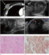
A 52-year-old man visited our emergency center for stroke and was hospitalized in neurology department. During hospital day, he complained of a palpable mass on the right breast that had been for 4 years and gradually increased in size. He had no family history of breast cancer and had a history of a rib fracture due to bicycle accident. After fracture, a hard mass was presented on the right breast, around the site, gradually increasing in size. Contrast-enhanced chest CT was performed with suspicion of bone related disease. CT image revealed a 3.3 cmsized, oval shaped, microlobulated marginated, mild enhancing soft tissue density mass in outer portion of right breast. It closely abutted right pectoralis major muscle and overlying skin was thickened (Fig. 1A). There was no significant mediastinal or axillary lymphadenopathy. Additional ultrasound showed a 3.5cm-sized oval shaped, microlobulated marginated heterogeneous hypoechoic mass with calcifications in the mass at palpable site of right breast outer portion. Associated findings including internal vascularity and skin thickening were also observed (Fig. 1B–D). Mammography and MR was not performed. Given the clinical and radiologic appearance of the mass, we classified the mass as suspicious malignancy (category 4B) under the Breast Imaging Reporting and Data System (BIRADS). Ultrasound-guided core needle biopsy with 14G needle for three times was performed. On microscopic examination, tumor cells showed cytologically bland nuclei and abundant cytoplasm with indistinct cell borders (Fig. 1E). The immunohistochemistry revealed diffuse positive reaction for S100 protein of the tumor cells (Fig. 1F). The patient transferred to another hospital and underwent surgical removal of the lesion. On gross examination, the tumor appeared well-circumscribed homogeneous yellowish mass without necrosis measuring 3.2 × 2.4 × 4.0 cm. Microscopic findings were GCTs consistent with core needle biopsy findings. One year after the surgery, there was no demonstrable recurrent tumor on right breast on the follow-up sonography.
DISCUSSION
GCT is an uncommon breast tumor. It was first described by Abrikossoff (5). Initially, it was thought to originate from striated muscle cells, so called them myoblastomas (1). The histogenesis of this lesion has still controversy, but the most widely accepted theory has been that of a schwann cell origin, because of GCTs' characteristic histological features and specific immunohistological staining for S-100 protein and neuron-specific enolase (13).
GCT can occur in any visceral and cutaneous site of body, most commonly in oral cavity. It has been reported patients with GCT of the breast are usually middle-aged, premenopausal women, most commonly in premenopausal African American women (1). Direct associations with estrogen and progesterone have not been documented (6). Only 5–8% of all GCTs occur in the breast, approximately 1 of every 1000 breast tumors, with a male-female ratio of 1:9 (23). Male patients account for 6.6% of all GCTs of the breast (4). We identified three cases of male breast GCT in Korea, two cases from the electronic database MEDLINE and one case in the Korean radiologic literature.
Clinically, GCTs manifest as unilateral, solitary, firm or hard painless mass (2). In the contrast of breast carcinoma, which occur more commonly in upper outer quadrant, GCTs is known to occur more frequently in the upper inner quadrant of the breast, and this distribution appears to correspond to the cutaneous sensory distribution of the supraclavicular nerve (23). Retraction of overlying skin, ulceration, fixation to pectoralis fascia, and nipple inversion can also accompany, thereby mimicking breast carcinoma (126). On our case, the palpable mass was developed in outer portion, but no definite retraction or ulceration of skin or nipple inversion was observed on physical examination.
The mammographic appearance of GCT is variable, from benign looking mass as a round, circumscribed mass to an indistinct or spiculated mass, indistinguishable from carcinoma (23). A GCT can appear as a new lesion or as a mass that enlarges over time (1). Microcalcifications are not common (1236).
Although CT scans had not been considered the primary method to evaluate specific breast lesions, incidental breast lesions on routine chest CT are increasingly being encountered. In our case, axial contrast-enhanced CT image shows approximately 3.3 cm sized, oval shaped microlobulated soft tissue density mass in right breast. Moreover, the mass demonstrates mild enhancement which is corresponding to arterial enhancement of malignant breast lesions on dynamic study (78). Other associated CT features including overlying skin thickening and close contact with right pectoralis major muscle also suggested the possibility of malignancy.
There is no generally acknowledged CT features of GCT of breast. According to some reports, CT images of GCT of the subcutis show subcutaneous mass with an ill-defined margin and isodense with the surrounding muscle (9). Microlobulated margin and isodensity of our case would correspond to the characteristics of GCT, in this regard.
The sonographic appearance of GCT is also variable, including a variable echotexture; circumscribed, angular, or spiculated margins; acoustics ranging from posterior shadowing to increase through transmission; and hypervascularity. Many GCTs would have a surrounding hyperechoic halo or partial hyperechoigenicity (12). Yang et al. (1) reported this hyperechogenicity may reflect the infiltrative growth pattern of this tumor in part and may be related to the heterogeneous cell origin of this lesion (perineural or Schwann cell differentiation).
On our case, the breast sonography demonstrated suspicious features of malignancy, including microlobulated margin, heterogeneous hypoechogenicity, and calcifications in the mass with internal vascularity and skin thickening. We classified the mass as category 4B (BIRADS). Notably, the mass showed no hyperechoic halo which is relatively common in GCT. Instead, calcifications in the mass was seen which is more common in breast carcinoma than in GCT. These sonographic features suggested suspicious malignancy.
For these variable radiologic features of GCT, core needle biopsy is required to distinguish GCTs from carcinoma. Microscopically, the tumor cells are arranged in nests or solid sheets, sometimes infiltrating deeply in the collagenous stroma. The most characteristic cytological feature of GCT is polygonal cells with abundant eosinophilic and finely granular cytoplasm (10). These cells show granular cytoplasm and strong cytoplasmatic and nuclear staining for the S-100 protein (4). In our case, the tumor is composed of polygonal cells with eosinophilic granular cells with diffuse immunoreactivity for S100 protein. Microscopically, it was typical benign GCT.
The majority of GCTs is benign and management depends on the clinical presentation, including follow-up imaging after biopsy, local or wide excision (2). Approximately 2–8% of GCTs reported recurrence after local excision when the resection margin is free from tumor infiltration. The overall prognosis is good, even if in recurrent cases (4). In our case, The patient transferred to another hospital and underwent surgical removal of the lesion. One year after the surgery, there was no demonstrable recurrent tumor on right breast on the follow-up sonography.
We describe a case of GCT of the breast with CT, sonography, and pathology. GCT is very rare benign tumor in breast, especially in male patient. Clinically, it usually manifests as palpable mass and imaging features on CT and sonography would mimic breast malignancy. But the clinical behavior and management of GCT is much more different from breast cancer. For the male patient with palpable mass in breast, benign GCT should be included in the differential diagnoses of heterogeneous solid lesions mimicking malignancy. We expect image findings of our case will be helpful for differential diagnosis and appropriate management.
 XML Download