Journal List > Korean J Radiol > v.19(6) > 1105326

Kang, Kim, Park, and Baek: Age of Data in Contemporary Research Articles Published in Representative General Radiology Journals

Abstract

Objective

To analyze and compare the age of data in contemporary research articles published in representative general radiology journals.

Materials and Methods

We searched for articles reporting original research studies analyzing patient data that were published in the print issues of the Korean Journal of Radiology (KJR), European Radiology (ER), and Radiology in 2017. Eligible articles were reviewed to extract data collection period (time from first patient recruitment to last patient follow-up) and age of data (time between data collection end and publication). The journals were compared in terms of the proportion of articles reporting the data collection period to the level of calendar month and regarding the age of data.

Results

There were 50, 492, and 254 eligible articles in KJR, ER, and Radiology, respectively. Of these, 44 (88%; 95% confidence interval [CI]: 75.8–94.8%), 359 (73%; 95% CI: 68.9–76.7%), and 211 (83.1%; 95% CI: 78–87.2%) articles, respectively, provided enough details of data collection period, revealing a significant difference between ER and Radiology (p = 0.002). The age of data was significantly greater in KJR (median age: 826 days; range: 299–2843 days) than in ER (median age: 570 days; range: 56–4742 days; p < 0.001) and Radiology (median age: 618; range: 75–4271 days; p < 0.001).

Conclusion

Korean Journal of Radiology did not fall behind ER or Radiology in reporting of data collection period, but showed a significantly greater age of data than ER and Radiology, suggesting that KJR should take measures to improve the timeliness of its data.

INTRODUCTION

The purpose of scientific journals is to share new information and novel discoveries with professionals of the relevant specialty. This is critical in medicine, where the application of new knowledge from journals advances clinical practice. For this reason, the timeliness of research data is essential; indeed, some journals provide submitting authors with specific guidelines in this regard (1). Moreover, in radiology, data timeliness may be especially important since the field relies heavily on new technology particularly digital technology (234567), which develops and changes faster than in other disciplines. Thus, the timeliness of data in radiology journals may be an important indicator of quality or impact, and the present study analyzed and compared several representative general radiology journals in terms of the age of the data published therein.

MATERIALS AND METHODS

This study did not require an Institutional Review Board approval.

Study Selection

We analyzed the Korean Journal of Radiology (KJR), European Radiology (ER), and Radiology, which have the highest impact factors among general radiology journals published in Asia, Europe, and America, respectively, according to the 2017 Journal Citation Reports (Clarivate Analytics, Philadelphia, PA, USA). We hand-searched the print issues of the journals published in 2017 (6 issues of KJR, 12 issues of ER, and 12 issues of Radiology) to find reports of original research studies that had collected and analyzed patient data. Experimental studies involving animals, phantoms, or other laboratory conditions were excluded. The literature was screened by two board-certified radiologists, both of whom were proficient in reviewing radiology research articles.

Data Extraction

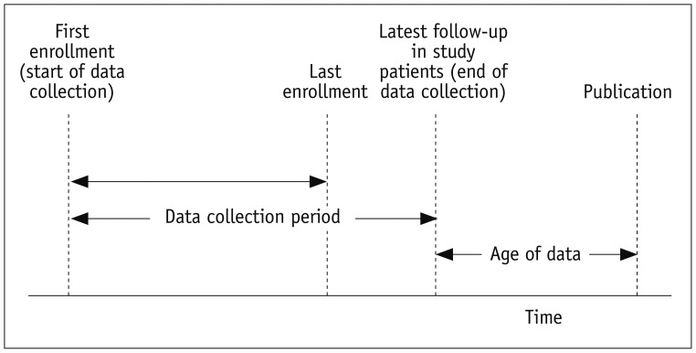
Firstly, the eligible articles were reviewed to determine whether they had stated the start and end of the data collection period to the level of the calendar month (Fig. 1). The data collection period was defined as the time from recruitment of the first patient to the latest follow-up of the study patients, as defined elsewhere (Fig. 1) (8). We considered month, but not date, as it is rare for published radiology research studies to report the data collection period to the level of date. Next, we made a universal assumption that the start and end dates of the data collection period fell on the first and last days, respectively, of the reported months. For example, if the data collection period ranged from January 2011 to April 2015, the start and end dates were assumed to be January 01, 2011 and April 30, 2015, respectively. As most studies only report the shortest and longest follow-up durations, rather than the exact follow-up periods of each study participant, the exact time of each patient's latest follow-up is rarely given. Therefore, we determined the time of the latest follow-up by 1) assuming that the earliest study enrollee had the longest follow-up and that the last enrollee had the shortest follow-up, 2) calculating two time points by adding the longest and shortest follow-up lengths to the start and end, respectively, of enrollment, and 3) choosing the later of these two time points. This method gave a reasonable estimate of the latest follow-up time. Secondly, the type of study design was determined (retrospective, prospective, or unclear). Thirdly, the exact date of publication, according to the time at which the full-text article became available online, was recorded by referring to the PubMed data of ER and Radiology, both of which use electronic publication ahead of print, and through contact with the editorial office of KJR, which does not use electronic publication ahead of print. Two board-certified radiologists analyzed the eligible articles to extract data. When there was ambiguity, a third reviewer experienced in the relevant methodology was invited.

Outcome Measures and Statistical Analysis

The proportion of articles that revealed the data collection period to the level of the calendar month was calculated, along with its 95% confidence interval (CI). The proportion was compared between journals in a pairwise manner using Fisher's exact test: KJR vs. ER, KJR vs. Radiology, and ER vs. Radiology. In articles that revealed the data collection period, the age of the research data was calculated as the time between the end of the data collection period and the date of publication (Fig. 1). The distribution of the age of data was checked using a histogram and the Kolmogorov-Smirnov test, and appropriate summary statistics were obtained. Next, the age of the data was compared between journals in the aforementioned pairwise manner using the Wilcoxon rank-sum test. This statistical comparison was performed across all relevant articles, as well as separately for prospective and retrospective studies. The threshold p value for statistical significance was lowered to 0.017 (Bonferroni adjustment) to maintain the overall alpha at 5% after the three pairwise comparisons were made. Statistical analyses were performed using SPSS Statistics for Windows, version 21.0 (IBM Corp., Armonk, NY, USA).

RESULTS

In total, 50 KJR (9101112131415161718192021222324252627282930313233343536373839404142434445464748495051525354565758), 492 ER, and 254 Radiology articles reported original research studies analyzing patient data. Of these, 44 (88%; 95% CI: 75.8–94.8%) (910121314151617181920212324262728293031323435363738394041424344474849505152535455565758), 359 (73%; 95% CI: 68.9–76.7%), and 211 (83.1%; 95% CI: 78–87.2%) articles, respectively, revealed the start and end of data collection to the level of calendar month. The point estimate value of this proportion was slightly larger in KJR than in ER and Radiology, although the difference was not statistically significant. In contrast, ER demonstrated a significantly lower proportion than Radiology in this regard (Table 1). Further breakdowns according to study type are also presented in Table 1, and the separate results from prospective and retrospective studies were mostly consistent with the overall results.
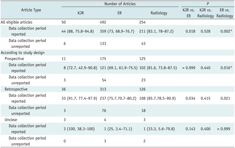
The age of data (the time between the end of the data collection period and the date of publication) was skewed to the left in all three journals (Fig. 2) and was significantly larger in KJR (median age: 826 days across all relevant articles) than in ER (median age, 570 days) and Radiology (median age: 618 days) (Table 2). The difference was more pronounced when retrospective studies were considered separately (Table 2).
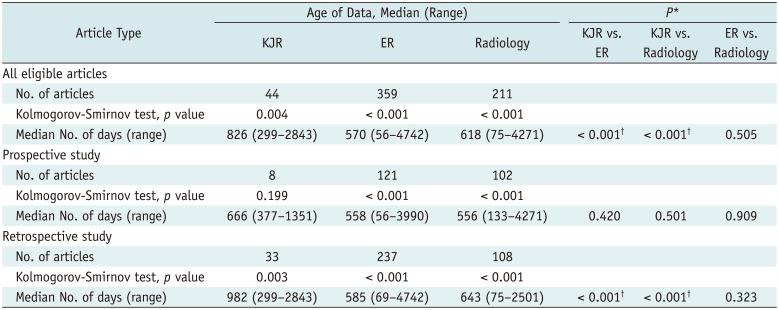
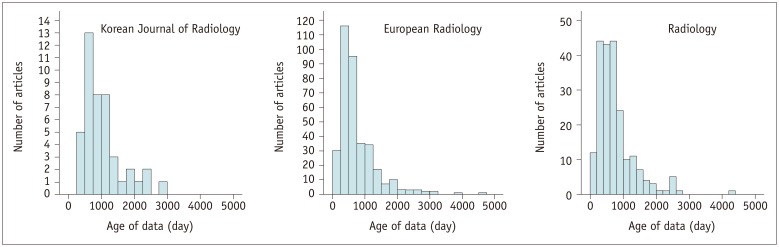
DISCUSSION

Korean Journal of Radiology seemed not to fall behind ER and Radiology regarding the proportion of articles that reported the data collection period, although the results of prospective studies may be inconclusive because too few relevant articles were published in KJR (11 articles). Nonetheless, this result may indicate that the journal has good quality control in the peer review and editorial processes. However, the age of data was significantly greater in KJR than in ER (approximately 8.5-month difference in median age) and Radiology (approximately 7-month difference in median age). Furthermore, the greater age of the data was more pronounced when retrospective studies were considered separately (approximately 13 months older than in ER and 11 months older than in Radiology). It is likely that the KJR contains older data because authors generally submit their manuscripts to higher-ranked journals first, descending the ranks of journals if their manuscript is rejected. As a result, lower-ranked journals would naturally contain older data. The journal impact factors of KJR, ER, and Radiology for 2017, according to the Journal Citation Reports (Clarivate Analytics), were 3.072, 4.027, and 7.469, respectively. This would explain why KJR differs from ER or Radiology in this regard.
However, there was no significant difference in the age of data between ER and Radiology, despite the apparent difference in journal impact factor, perhaps because ER publishes accepted articles more swiftly than Radiology in electronic publication format ahead of print publication. Specifically, in the articles analyzed in the present study, the median interval between initial electronic publication and official assignment to a monthly print issue was 225 days (range: 130–316 days) for ER and 138 days (range: 39–244 days) for Radiology.
Korean Journal of Radiology recently assessed the quality of its research articles in terms of conformity to the Standards for Reporting of Diagnostic Accuracy Studies (STARD) 2015 guidelines (59) and the adequacy reporting reliability analysis for diagnostic tests (60). The current study revealed another area in which KJR could improve its quality and impact, and we would even suggest some specific measures. Firstly, because the age of data in the KJR differed more markedly from that in ER and Radiology in retrospective studies than in prospective studies, the journal could encourage authors to make data as recent as possible by updating study data in the revision process. Such an update would be possible in the case of retrospective studies, although it is likely infeasible in most prospective studies. Secondly, the journal could further shorten the time from initial submission to publication by ensuring more rapid review, allowing electronic publication before print, and publishing monthly in a smaller volume. Indeed, bimonthly publication delays those articles that were early accepted in the 2-month cycle period.
This study had several limitations. Firstly, as studies generally do not report each individual's exact follow-up duration, we assumed that the earliest study enrollee had the longest follow-up and the latest enrollee had the shortest follow-up; this may not always have been the case. However, we believe that this approach gave a reasonable estimate of the data collection period and was sufficient to analyze the macroscopic, between-journal difference. Secondly, one published study (8) defined the age of data slightly differently from the present study as the time from the mid-point of the data collection period to the publication date. This previous study analyzed prospective randomized trials, wherein their definition fit better. However, this definition would have been inappropriate in our study, wherein the majority of studies analyzed were retrospective. In retrospective studies, a data collection time extending further into the past may be more beneficial. However, for the purposes of the present study, it may have penalized the results, as the data age became greater. Thirdly, knowledge of the details of any rejections by other journals (number of rejections, by what journals, etc.) before submission to KJR would have been helpful in further understanding the greater age of KJR data. However, we empirically found that it was difficult to collect such “sensitive” information with consistency.
In conclusion, KJR did not fall behind ER or Radiology with regard to the proportion of articles that reported the age of data. However, the age of data was significantly greater in KJR than in ER and Radiology, suggesting that the journal should introduce some measures to improve the timeliness of data that it publishes.

References

1. Instructions for Authors. JAMA Web site. 2018. 8. 01. Accessed August 24, 2018. https://jamanetwork.com/journals/jama/pages/instructions-for-authors.
2. Ahn S, Park SH, Lee KH. How to demonstrate similarity by using noninferiority and equivalence statistical testing in radiology research. Radiology. 2013; 267:328–338. PMID: 23610094.
crossref
3. Lee JG, Jun S, Cho YW, Lee H, Kim GB, Seo JB, et al. Deep Learning in medical imaging: general overview. Korean J Radiol. 2017; 18:570–584. PMID: 28670152.
crossref
4. Park SH, Han K. Methodologic guide for evaluating clinical performance and effect of artificial intelligence technology for medical diagnosis and prediction. Radiology. 2018; 286:800–809. PMID: 29309734.
5. Park SH, Kressel HY. Connecting technological innovation in artificial intelligence to real-world medical practice through rigorous clinical validation: what peer-reviewed medical journals could do. J Korean Med Sci. 2018; 33:e152. PMID: 29805337.
crossref
6. Kim GB, Lee S, Kim H, Yang DH, Kim YH, Kyung YS, et al. Three-dimensional printing: basic principles and applications in medicine and radiology. Korean J Radiol. 2016; 17:182–197. PMID: 26957903.
crossref
7. Kim M, Kim HS. Emerging techniques in brain tumor imaging: what radiologists need to know. Korean J Radiol. 2016; 17:598–619. PMID: 27587949.
crossref
8. Welsh J, Lu Y, Dhruva SS, Bikdeli B, Desai NR, Benchetrit L, et al. Age of data at the time of publication of contemporary clinical trials. JAMA Network Open. 2018; 1:e181065.
crossref
9. Ahn SJ, Lee JM, Chang W, Lee SM, Kang HJ, Yang H, et al. Prospective validation of intra- and interobserver reproducibility of a new point shear wave elastographic technique for assessing liver stiffness in patients with chronic liver disease. Korean J Radiol. 2017; 18:926–935. PMID: 29089825.
crossref
10. Aslaner R, Pekcevik Y, Sahin H, Toka O. Variations in the origin of inferior phrenic arteries and their relationship to celiac axis variations on CT angiography. Korean J Radiol. 2017; 18:336–344. PMID: 28246513.
crossref
11. Azad R, Tayal M, Azad S, Sharma G, Srivastava RK. Qualitative and quantitative comparison of contrast-enhanced fluid-attenuated inversion recovery, magnetization transfer spin echo, and fat-saturation T1-weighted sequences in infectious meningitis. Korean J Radiol. 2017; 18:973–982. PMID: 29089830.
crossref
12. Bae YJ, Choi BS, Jung C, Yoon YH, Sunwoo L, Bae HJ, et al. Differentiation of deep subcortical infarction using high-resolution vessel wall MR imaging of middle cerebral artery. Korean J Radiol. 2017; 18:964–972. PMID: 29089829.
crossref
13. Bae YJ, Choi BS, Lee KM, Yoon YH, Sunwoo L, Jung C, et al. Efficacy of maximum intensity projection of contrast-enhanced 3D turbo-spin echo imaging with improved motion-sensitized driven-equilibrium preparation in the detection of brain metastases. Korean J Radiol. 2017; 18:699–709. PMID: 28670165.
crossref
14. Becker AS, Perucho JA, Wurnig MC, Boss A, Ghafoor S, Khong PL, et al. Assessment of cervical cancer with a parameter-free intravoxel incoherent motion imaging algorithm. Korean J Radiol. 2017; 18:510–518. PMID: 28458603.
crossref
15. Chae HD, Lee JY, Jang JY, Chang JH, Kang J, Kang MJ, et al. Photoacoustic imaging for differential diagnosis of benign polyps versus malignant polyps of the gallbladder: a preliminary study. Korean J Radiol. 2017; 18:821–827. PMID: 28860899.
crossref
16. Chan WY, Chong LR. Anatomical variants of lister's tubercle: a new morphological classification based on magnetic resonance imaging. Korean J Radiol. 2017; 18:957–963. PMID: 29089828.
crossref
17. Chen SQ, Huang M, Shen YY, Liu CL, Xu CX. Abbreviated MRI protocols for detecting breast cancer in women with dense breasts. Korean J Radiol. 2017; 18:470–475. PMID: 28458599.
crossref
18. Cheng Y, Huang LX, Zhang L, Ma M, Xie SS, Ji Q, et al. Longitudinal intrinsic brain activity changes in cirrhotic patients before and one month after liver transplantation. Korean J Radiol. 2017; 18:370–377. PMID: 28246517.
crossref
19. Choi IY, Park SH, Park SH, Yu CS, Yoon YS, Lee JL, et al. CT enterography for surveillance of anastomotic recurrence within 12 months of bowel resection in patients with Crohn's disease: an observational study using an 8-year registry. Korean J Radiol. 2017; 18:906–914. PMID: 29089823.
crossref
20. Choi TW, Lee JM, Lee DH, Lee JH, Yu SJ, Kim YJ, et al. Percutaneous dual-wwitching monopolar radiofrequency ablation using a separable clustered electrode: a preliminary study. Korean J Radiol. 2017; 18:799–808. PMID: 28860897.
21. Chung HW, Ko SM, Hwang HK, So Y, Yi JG, Lee EJ. Diagnostic performance of coronary CT angiography, stress dual-energy CT perfusion, and stress perfusion single-photon emission computed tomography for coronary artery disease: comparison with combined invasive coronary angiography and stress perfusion cardiac MRI. Korean J Radiol. 2017; 18:476–486. PMID: 28458600.
crossref
22. Feng R, Tong J, Liu X, Zhao Y, Zhang L. High-pitch coronary CT angiography at 70 kVp adopting a protocol of low injection speed and low volume of contrast medium. Korean J Radiol. 2017; 18:763–772. PMID: 28860894.
crossref
23. Goo HW, Allmendinger T. Combined electrocardiography- and respiratory-triggered CT of the lung to reduce respiratory misregistration artifacts between imaging slabs in free-breathing children: initial experience. Korean J Radiol. 2017; 18:860–866. PMID: 28860904.
crossref
24. Huh J, Kim KJ, Park SH, Park SH, Yang SK, Ye BD, et al. Diffusion-weighted MR enterography to monitor bowel inflammation after medical therapy in Crohn's disease: a prospective longitudinal study. Korean J Radiol. 2017; 18:162–172. PMID: 28096726.
crossref
25. Hwang JS, Lee H, Lee B, Lee SJ, Jou SS, Lim HK, et al. Estimation of diastolic filling pressure with cardiac CT in comparison with echocardiography using tissue Doppler imaging: determination of optimal CT reconstruction parameters. Korean J Radiol. 2017; 18:632–642. PMID: 28670158.
crossref
26. Hyun D, Park KB, Cho SK, Park HS, Shin SW, Choo SW, et al. Portal vein stenting for delayed jejunal varix bleeding associated with portal venous occlusion after hepatobiliary and pancreatic surgery. Korean J Radiol. 2017; 18:828–834. PMID: 28860900.
crossref
27. Jang J, Kim TW, Hwang EJ, Choi HS, Koo J, Shin YS, et al. Assessment of arterial wall enhancement for differentiation of parent artery disease from small artery disease: comparison between histogram analysis and visual analysis on 3-dimensional contrast-enhanced T1-weighted turbo spin echo MR images at 3T. Korean J Radiol. 2017; 18:383–391. PMID: 28246519.
crossref
28. Jeon TY, Kim JH, Lee J, Yoo SY, Hwang SM, Lee M. Value of repeat brain MRI in children with focal epilepsy and negative findings on initial MRI. Korean J Radiol. 2017; 18:729–738. PMID: 28670168.
crossref
29. Kang EJ, Lee KN, Choi WJ, Kim YD, Shin KM, Lim JK, et al. Left ventricular functional parameters and geometric patterns in Korean adults on coronary CT angiography with a 320-detector-row CT scanner. Korean J Radiol. 2017; 18:664–673. PMID: 28670161.
crossref
30. Kang TW, Lee MW, Song KD, Kim M, Kim SS, Kim SH, et al. Added value of contrast-enhanced ultrasound on biopsies of focal hepatic lesions invisible on fusion imaging guidance. Korean J Radiol. 2017; 18:152–161. PMID: 28096725.
crossref
31. Kang Y, Lee GY, Lee JW, Lee E, Kim B, Kim SJ, et al. Texture analysis of torn rotator cuff on preoperative magnetic resonance arthrography as a predictor of postoperative tendon status. Korean J Radiol. 2017; 18:691–698. PMID: 28670164.
crossref
32. Kim B, Yoon DY, Seo YL, Park MW, Kwon KH, Rho YS, et al. Value of the post-operative CT in predicting delayed flap failures following head and neck cancer surgery. Korean J Radiol. 2017; 18:536–542. PMID: 28458606.
crossref
33. Kim DJ, Park MK, Jung DE, Kang JH, Kim BM. Radiation dose reduction without compromise to image quality by alterations of filtration and focal spot size in cerebral angiography. Korean J Radiol. 2017; 18:722–728. PMID: 28670167.
crossref
34. Kim GE, Shin SS, Kim JW, Heo SH, Lim HS, Jun CH, et al. Incidental, small (< 3 cm), unilocular, pancreatic cysts: factors that predict lesion progression during imaging surveillance. Korean J Radiol. 2017; 18:915–925. PMID: 29089824.
35. Kim HJ, Bang JI, Kim JY, Moon JH, So Y, Lee WW. Novel application of quantitative single-photon emission computed tomography/computed tomography to predict early response to methimazole in Graves' disease. Korean J Radiol. 2017; 18:543–550. PMID: 28458607.
crossref
36. Kim JS, Kim HJ, Hong SM, Park SH, Lee JS, Kim AY, et al. Post-ischemic bowel stricture: CT features in eight cases. Korean J Radiol. 2017; 18:936–945. PMID: 29089826.
crossref
37. Kim SK, Lee KA, Sauk S, Korenblat K. Comparison of transjugular intrahepatic portosystemic shunt with covered stent and balloon-occluded retrograde transvenous obliteration in managing isolated gastric varices. Korean J Radiol. 2017; 18:345–354. PMID: 28246514.
crossref
38. Kim SS, Jin GY, Li YZ, Lee JE, Shin HS. CT quantification of lungs and airways in normal Korean subjects. Korean J Radiol. 2017; 18:739–748. PMID: 28670169.
crossref
39. Kim YJ, Lee EH, Jun JK, Shin DR, Park YM, Kim HW, et al. Analysis of participant factors that affect the diagnostic performance of screening mammography: a report of the alliance for breast cancer screening in Korea. Korean J Radiol. 2017; 18:624–631. PMID: 28670157.
crossref
40. Kim YP, Haam SJ, Lee S, Lee GD, Joo SM, Yum TJ, et al. Effectiveness of ambulatory tru-close thoracic vent for the outpatient management of pneumothorax: a prospective pilot study. Korean J Radiol. 2017; 18:519–525. PMID: 28458604.
crossref
41. Lee GM, Kim YR, Ryu JH, Kim TH, Cho EY, Lee YH, et al. Quantitative measurement of hepatic fibrosis with gadoxetic acid-enhanced magnetic resonance imaging in patients with chronic hepatitis B infection: a comparative study on aspartate aminotransferase to Platelet Ratio Index and Fibrosis-4 Index. Korean J Radiol. 2017; 18:444–451. PMID: 28458596.
crossref
42. Lee JS, Kim SH, Im SA, Kim MA, Han JK. Human epidermal growth factor receptor 2 expression in unresectable gastric cancers: relationship with CT characteristics. Korean J Radiol. 2017; 18:809–820. PMID: 28860898.
crossref
43. Lee JW, Jeong YJ, Lee G, Lee NK, Lee HW, Kim JY, et al. Predictive value of cardiac magnetic resonance imaging-derived myocardial strain for poor outcomes in patients with acute myocarditis. Korean J Radiol. 2017; 18:643–654. PMID: 28670159.
crossref
44. Lee MS, Moon MH, Woo H, Sung CK, Jeon HW, Lee TS. Ruptured corpus luteal cyst: prediction of clinical outcomes with CT. Korean J Radiol. 2017; 18:607–614. PMID: 28670155.
crossref
45. Min ZG, Niu C, Zhang QL, Zhang M, Qian YC. Optimal factors of diffusion tensor imaging predicting corticospinal tract injury in patients with brain tumors. Korean J Radiol. 2017; 18:844–851. PMID: 28860902.
crossref
46. Nam HY, Jun S, Pak K, Kim IJ. Concurrent low brain and high liver uptake on FDG PET are associated with cardiovascular risk factors. Korean J Radiol. 2017; 18:392–401. PMID: 28246520.
crossref
47. Park HJ, Kim HJ, Park SH, Lee JS, Kim AY, Ha HK. Gastrointestinal involvement of recurrent renal cell carcinoma: CT findings and clinicopathologic features. Korean J Radiol. 2017; 18:452–460. PMID: 28458597.
crossref
48. Park JE, Park B, Kim SJ, Kim HS, Choi CG, Jung SC, et al. Improved diagnostic accuracy of Alzheimer's disease by combining regional cortical thickness and default mode network functional connectivity: validated in the Alzheimer's disease neuroimaging initiative set. Korean J Radiol. 2017; 18:983–991. PMID: 29089831.
crossref
49. Seo M, Ryu JK, Jahng GH, Sohn YM, Rhee SJ, Oh JH, et al. Estimation of T2* relaxation time of breast cancer: correlation with clinical, imaging and pathological features. Korean J Radiol. 2017; 18:238–248. PMID: 28096732.
crossref
50. Sung J, Jee WH, Jung JY, Jang J, Kim JS, Kim YH, et al. Diagnosis of nerve root compromise of the lumbar spine: evaluation of the performance of three-dimensional isotropic T2-weighted turbo spin-echo SPACE sequence at 3T. Korean J Radiol. 2017; 18:249–259. PMID: 28096733.
crossref
51. Tan Y, Zhou J, Zhou Y, Yang X, Yang J, Chen Y. Characteristics detected on computed tomography angiography predict coronary artery plaque progression in non-culprit lesions. Korean J Radiol. 2017; 18:487–497. PMID: 28458601.
crossref
52. Tang YL, Zhang XM, Yang ZG, Huang YC, Chen TW, Chen YL, et al. The blood oxygenation T2* values of resectable esophageal squamous cell carcinomas as measured by 3T magnetic resonance imaging: association with tumor stage. Korean J Radiol. 2017; 18:674–681. PMID: 28670162.
53. Xie S, Li Q, Cheng Y, Zhang Y, Zhuo Z, Zhao G, et al. Impact of liver fibrosis and fatty liver on T1rho measurements: a prospective study. Korean J Radiol. 2017; 18:898–905. PMID: 29089822.
crossref
54. Yang SY, Lee KS, Cha MJ, Kim TJ, Kim TS, Yoon HJ. Chest CT features of cystic fibrosis in Korea: comparison with non-cystic fibrosis diseases. Korean J Radiol. 2017; 18:260–267. PMID: 28096734.
crossref
55. Yoo H, Lee JM, Yoon JH, Kang HJ, Lee SM, Yang HK, et al. T2* mapping from multi-echo dixon sequence on gadoxetic acid-enhanced magnetic resonance imaging for the hepatic fat quantification: can It be used for hepatic function assessment? Korean J Radiol. 2017; 18:682–690. PMID: 28670163.
crossref
56. Yu M, Li Y, Li W, Lu Z, Wei M, Zhang J. Calcification remodeling index characterized by Cardiac CT as a novel parameter to predict the use of rotational atherectomy for coronary intervention of lesions with moderate to severe calcification. Korean J Radiol. 2017; 18:753–762. PMID: 28860893.
crossref
57. Yu M, Zhang Y, Li Y, Li M, Li W, Zhang J. Assessment of myocardial bridge by cardiac CT: intracoronary transluminal attenuation gradient derived from diastolic phase predicts systolic compression. Korean J Radiol. 2017; 18:655–663. PMID: 28670160.
crossref
58. Zhang Y, Huang QH, Fang Y, Yang P, Xu Y, Hong B, et al. A novel flow diverter (tubridge) for the treatment of recurrent aneurysms: a single-center experience. Korean J Radiol. 2017; 18:852–859. PMID: 28860903.
crossref
59. Choi YJ, Chung MS, Koo HJ, Park JE, Yoon HM, Park SH. Does the reporting quality of diagnostic test accuracy studies, as defined by STARD 2015, affect citation? Korean J Radiol. 2016; 17:706–714. PMID: 27587959.
crossref
60. Park JE, Han K, Sung YS, Chung MS, Koo HJ, Yoon HM, et al. Selection and reporting of statistical methods to assess reliability of a diagnostic test: conformity to recommended methods in a peer-reviewed journal. Korean J Radiol. 2017; 18:888–897. PMID: 29089821.
crossref
Fig. 1

Definitions of data collection period and age of data.

kjr-19-1172-g001
Fig. 2

Distribution of age of data.

kjr-19-1172-g002
Table 1

Articles Included in Study and Proportion of Articles Reporting Data Collection Period

kjr-19-1172-i001
Article Type Number of Articles P
KJR ER Radiology KJR vs. ER KJR vs. Radiology ER vs. Radiology
All eligible articles 50 492 254
 Data collection period reported 44 (88, 75.8–94.8) 359 (73, 68.9–76.7) 211 (83.1, 78–87.2) 0.018 0.528 0.002*
 Data collection period unreported 6 133 43
According to study design
 Prospective 11 175 125
  Data collection period reported 8 (72.7, 42.9–90.8) 121 (69.1, 61.9–75.5) 102 (81.6, 73.8–87.5) > 0.999 0.440 0.016*
  Data collection period unreported 3 54 23
 Retrospective 36 313 126
  Data collection period reported 33 (91.7, 77.4–97.9) 237 (75.7,70.7–80.2) 108 (85.7,78.5–90.9) 0.034 0.415 0.021
  Data collection period unreported 3 76 18
 Unclear 3 4 3
  Data collection period reported 3 (100, 38.3–100) 1 (25, 3.4–71.1) 1 (33.3, 5.6–79.8) 0.143 0.400 > 0.999
  Data collection period unreported 0 3 2

Data represent number of articles, with % of articles and corresponding 95% confidence intervals in parentheses. *Statistically significant after accounting for multiple comparisons (p < 0.017). ER = European Radiology, KJR = Korean Journal of Radiology

Table 2

Comparison of Age of Data between Journals

kjr-19-1172-i002
Article Type Age of Data, Median (Range) P*
KJR ER Radiology KJR vs. ER KJR vs. Radiology ER vs. Radiology
All eligible articles
 No. of articles 44 359 211
 Kolmogorov-Smirnov test, p value 0.004 < 0.001 < 0.001
 Median No. of days (range) 826 (299–2843) 570 (56–4742) 618 (75–4271) < 0.001 < 0.001 0.505
Prospective study
 No. of articles 8 121 102
 Kolmogorov-Smirnov test, p value 0.199 < 0.001 < 0.001
 Median No. of days (range) 666 (377–1351) 558 (56–3990) 556 (133–4271) 0.420 0.501 0.909
Retrospective study
 No. of articles 33 237 108
 Kolmogorov-Smirnov test, p value 0.003 < 0.001 < 0.001
 Median No. of days (range) 982 (299–2843) 585 (69–4742) 643 (75–2501) < 0.001 < 0.001 0.323

*Comparison of age of data between journals, Statistically significant after accounting for multiple comparisons (p < 0.017).

TOOLS
Similar articles