INTRODUCTION
MATERIALS AND METHODS
Patients
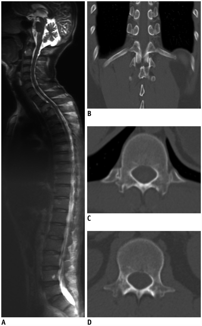 | Fig. 147-year-old woman with anomalous total number of vertebra.
A. Sagittal T2-weighted fast spin-echo magnetic resonance image of whole spine shows 25 presacral mobile vertebra. B, C. Coronal CT image (B) showing 20th vertebra, with paired ribs that are 3.8 cm or greater in length. Paired ribs originate from facet at pedicle of 20th vertebra on axial CT image (C). Therefore, 20th vertebra is morphologically thoracic vertebra. D. On axial CT image, 21st vertebra morphologically appears to be first lumbar vertebra. Thus, this patient has 25 presacral mobile vertebra with seven cervical, 13 thoracic, and five lumbar vertebra. CT = computed tomography
|
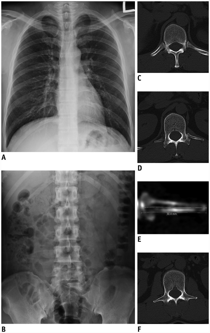 | Fig. 238-year-old man with TLTV.
A, B. Posteroanterior chest radiograph (A) and supine abdominal radiograph (B) demonstrate 24 presacral mobile vertebra. C. Axial CT image showing 19th vertebra, with paired ribs that are 3.8 cm or greater in length and originate from facet at pedicle. Therefore, 19th vertebra is morphologically thoracic vertebra. D. On axial CT image, 20th vertebra, which has short rib on left side and accessory ossification center on right side, is TLTV. E. On curved planar reformatted image of left rib of 20th vertebra, rib length is measured by drawing line at midpoint of rib width from proximal head of rib to distal body. Rib measures 30.4 mm and is classified as short rib. F. On axial CT image, 21st vertebra, exhibiting both fused transverse processes without articulating ribs, is identified as first lumbar vertebra. Thus, this patient has 24 presacral mobile vertebra with seven cervical, 12 thoracic, one TLTV, and four lumbar vertebra. TLTV = thoracolumbar transitional vertebra
|
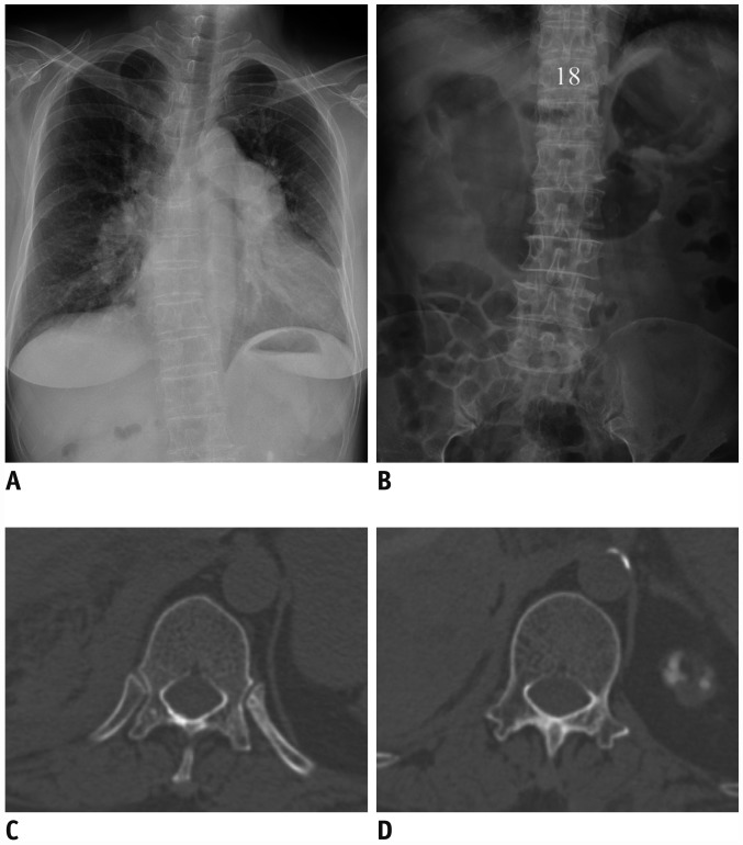 | Fig. 374-year-old woman with anomalous distribution of vertebra.
A, B. Posteroanterior chest radiograph (A) and supine abdominal radiograph (B) indicate presence of 24 presacral mobile vertebra. C. Axial CT image showing 18th vertebra, with paired ribs that are 3.8 cm or greater in length and originate from facet at pedicle. Therefore, 18th vertebra is morphologically thoracic vertebra. D. On axial CT image, 19th vertebra has both fused transverse processes and morphologically appears to be first lumbar vertebra. Thus, this patient has anomalous distribution of 24 presacral mobile vertebra with seven cervical, 11 thoracic, and six lumbar vertebra.
|
Spinal CT
Image Analysis
Establishment of the First Lumbar Vertebra in Spinal Variants: Morphologic Analysis of the Thoracolumbar Junction
Comparison of Spinal Enumeration Methods for Spinal Variants
RESULTS
Table 1
Characteristics of Study Patients with Spinal Variants
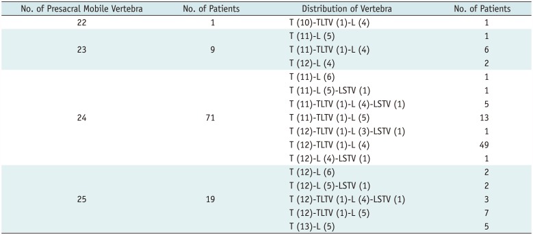
Table 2
Comparison of Methods for Correct Identification of First Lumbar Vertebra


 PDF
PDF ePub
ePub Citation
 Print
Print


 XML Download