Abstract
Successful repair of lacerated tendons must restore continuity of the tendon and should yield a strong tenorrhaphy. Mechanical strength of repair should be adequate to early postoperative motion and mobility. The optimal repair technique must be able to withstand the rigors of early motion and also must not interfere with tendon healing.
The relative strength of three suture methods of lacerated tendon were measured by mechanical disruption in effort to determine the strength of suture technique.
Fifty-four calcaneal tendons of 27 the New Zealand white rabbit were transected at mid portion and repaired with the three suture techniques: group 1, Kessler suture, group 2, Pennington’s modified-Kessler suture and group 3, augmented-Becker suture technique. Each group was composed of 18 calcaneal tendons. Nine rabbits were sacrified immediately after suture, nine in postoperative 2 weeks and nine in postoperative 4 weeks.
Six calcaneal tendons in each three experimental group were obtained immediately after suture, at postoperative 2 and 4 weeks respectively.
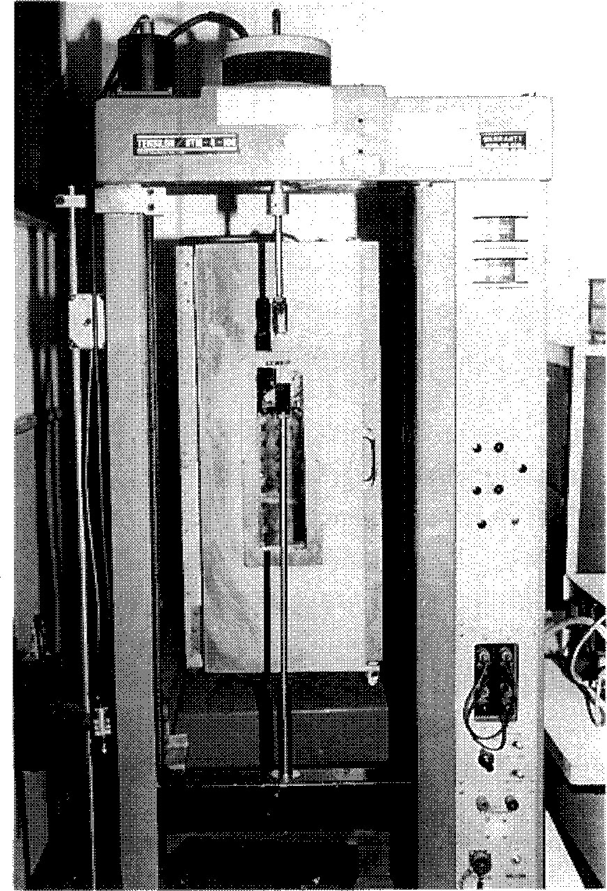
Tensile strength, maximum strength and modulus of elasticity of all experimental specimens were measured with Instron-UTM-4-100(Tyo-Baldiwin, Japan).
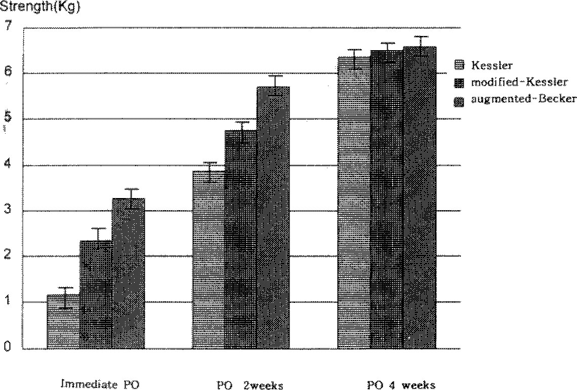
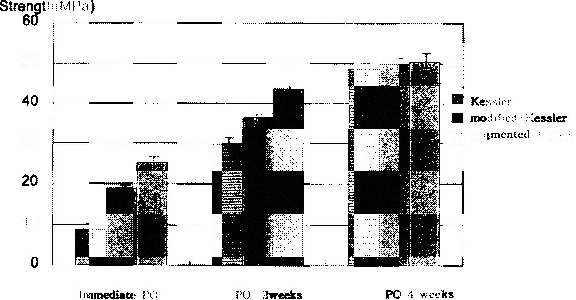
The results were evaluated statistically to compare the strengh of the three suture technique at three different periods. The tensile strength was predominantly strongest in augmented-Becker method among three suture technique at immediate suture, postoperative 2 weeks and 4 weeks respectively. The augmented Becker repair was strongest in maximum stress among Kessler and modified Kessler repair at immediate operation, postoperative 2 weeks and 4 weeks respectively.
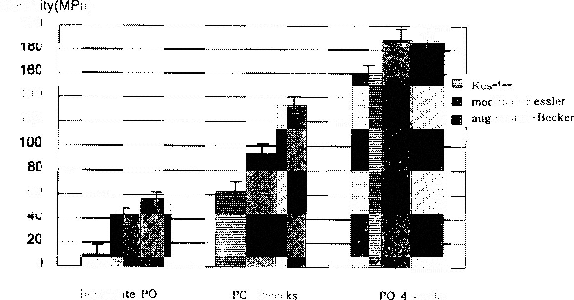
The augmented Becker repair was highest in modulus of elasticity than Kessler method and modified-Kessler method at immediate operation, postoperative 2 weeks and postoperative 4 weeks respectively. Tensile strength, maximum stress and modulus of elasticity were gradully increased from immediate operation to postoperative 4 weeks, but there were not statistically significance between experimental three suture methods at postoperative 4 weeks.
Go to : 
REFERENCES
1. Abrahams M. Mechanical behavior of tendon in vitro. Med & Bioi Eng. 1967; 5:433–443.
2. Abrahamsson S, Lundborg G, Lohmander S. Tendon healing in vivo. Scand J Plast Reconstr Surg. 1989; 12:199.
3. Becker H., Davidoff M. Elimination from the halsted peripheral suture. Hand. 1977; 9:306–311.
4. Becker H, Diegelmann RF. The influence of tension on intrinsic tendon fibroplasia. Orthop Rev. 1984; 13:153–9.
5. Carlstedt CA, Skagervall R. A model for computer-aided analysis of biomechanical properties of the plantaris longus tendon in the rabbit. J Biomechanics. 1986; 19-3:251–256.

6. Daniel P, Greenwald DP, Hong HZ, May J. Mechanical analysis of tendon suture techniques. J Hand Surg. 1994; 19(4):641–647.
7. Modd EM, Sigel B, Dunn MR. Localization of new cell formation in tendon healing by tritiated thymidine autoradiography. Surg Gynecol Obstet. 1966; 122:805–806.
8. Fung YCB. Elasticity of soft tissues in simple elongation. Am J Physiol. 1967; 213-216:1532–1544.

9. Gelberman RH. Flexor tendon healing and restoration of the gliding surface: An ultra-structural study in dogs. J Bone Joint Surg. 65-A:70–80. 1983.

10. Gordon L, Garrison JL, Cheng JC, Lin YK, Nathan RP, Levinsohn DG. Biomechanical analysis of a stepcut technique for flexor tendon repair. J Hand Surg. 1992; 17B:282–5.

11. Greenwald DP, Mass DP, Gottlieb LJ, Tuel R. Biomechanicasl analysis of intrinsic tendon healing in vitro and the effects of vitamins A and E. Plast Reconstr Surg. 1991; 87:925–930.
12. Hitchcock TF, Light TR, Bunch WH. The effect of immediate constrained digital motion on the strength of flexor tendon repairs in chickens. J Hand Surg. 1987; 12A:590–595.

13. Joseph T, Barmakian. Hua L, Steven M, Green. Martin A, Posner. Robert S. Casar : Comparision of a suture technique with the modified Kessler method: Resistance to gap formation. J hand Sur. 1994; 19A:777–781.
14. Kessler I, Nissim F. Primary repair without immobilization of flexor tendon division within the digital sheath. Acta Orthop Scand. 1969; 40:587–601.
15. Ketchum LD. Suture materials and suture thechnique used in tendon repair. Hand Clinic. 1985; 1:43.
16. Kim SK. Further evolution of the grasping technique for tendon repair. Ann Plast Surg. 1981; 7:113–119.

17. Leddy JP, Green DP. Flexor tendon’s acute injuries: Operative hand surgery. 1993. 3rd ed„. Churchill Li vingstone New York;p. 1823–1851.
18. Lundborg G. Experimental flexor tendon healing nutrition and intrinsic healing mechanisms. Hand,. 1976; 8:235–238.
19. Lesker PA. Histologic evidence of intrinsic flexor tendon repair in various experimental animals in vitro study. Clin Orthop,. 1984; 182:297–304.
20. Mass DP, Tuel R. Human flexor tendon repair process. J Hand Surg. 1989; 14A:64–71.
21. Mathews P, Richards H. Factors in the adherence of flexor tendon after repair. An experimental study in the rabbit. J Bone Joint Surg. 1976; 588:230–236.
22. Woo SL. A new methodology to determine the mechanical properties of ligaments at high strain rates. Biomechanical Engineering,. 1986; 108:365–367.
23. Pruitt DL, Manske PR. Fink В : Cyclic stress analysis of flexor tendon repair. Hand Surg,. 1991; 16A:701–707.
24. Rick P, William H, Seta J, Paul S. Brian В Clevelan OH Biomechanical evaluation and clinical evaluation of the epitenon-first technique of the flexor tendon repair. J Hand Surg,. 1995; 20A:261–266.
25. Russet J, Manske PR. Collagen synthesis during primary flexor tendon repair in vitro. J Orthop Res,. 1990; 8:13–20.
26. Sander WE. Advantages of “epitenon-first” suture placement technique in flexor tendon repair. Clin Orthop,. 280:198–199. 1992.
27. Sanjeevi R, Somanathan N, Ramaswamy D. A Viscoelastic model forcollagen fibers. Biomechanics. 1982; 15-3:1891–1983.
28. Slack C, Flint MH, Thompson BM. The influence of tension on intrinsic tendon fibroplasia. Orthop Rev. 13:153–159. 1984.
29. Urbaniak JR, Cahill JD, Mortenson RA. Tendon suturing methods. Analysis of tensile strength. In A.A.O.S symposium on tendon surgery in the hand. 1975. Mosby: St. Lous;p. 70–80.
30. Verdan С : Reparative surgery of flexor tendons in the digits. Tendon Surgery of the hand. 1979. Churchill Li vingstone;London:
31. Wade PJF, Mnir IFK, Hutcheon LL. Primary flexor tendon repair. The mechanical limitations of the modified Kessler technique. J Hand Surg. 1986; 11B:71–76.

32. Wray EC, Weeks PM. Experimental comparison of techniques of tendon repair. J Hand Surg. 1980; 5A:5144–5148.
Go to : 
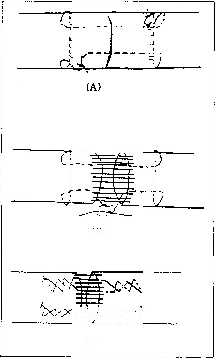 | Fig. 1Schematic drawing of tendon repair technique. A. Kessler technique with 4-0 nylon, B. Pennington’s modified Kessler technique with core stitch of 4-0 nylon and epitenon - first suture with 5-0 vicryl. C. Augmented - Becker repair with two rows of criss-crossing 4-0 nylon and epitenon - first suture with 5-0 vicryl. |
Table 1
Suture method, suture material and model setting of Instron Model -UTM-4-100(Toto-Baldiwin, Japan)
Table 2
Biomechanical properties of tendon repair with Kessler method.
Table 3
Biomechanical properties of tendon repair with modified-Kessler method.
Table 4
Biomechanical properties of tendon repair with augmented-Becker method.


 PDF
PDF ePub
ePub Citation
 Print
Print



 XML Download