

 PDF
PDF ePub
ePub Citation
 Print
Print


INTRODUCTION
Infarction of the recurrent artery of Heubner (RAH) is a rare complication following clipping of an anterior communicating artery (ACoA) aneurysm. The cause of the infarction is usually complication resulting from a subarachnoid hemorrhage (SAH), such as an operative maneuver or vasospasm. Bilateral RAH infarction following clipping of an ACoA aneurysm is relatively rare. Here, we report our experience with the territorial infarction of bilateral RAH following clipping of an ACoA aneurysm.
Go to : 
CASE REPORT
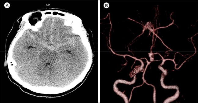
A 50-year-old woman presented to the emergency department with thunderclap headache and vomiting. She showed no neurologic deficit except for neck stiffness. Non-enhanced brain computed tomography (CT) showed a subarachnoid hemorrhage of Hunt-Hess Grade II and Fisher Grade III. Brain angiography CT revealed an aneurysm of the ACoA. The aneurysm was left dominant, directed inferiorly, and multi-lobulated (Fig. 1). On the transfemoral cerebral angiography (TFCA) performed pre-operatively, the right A1 segment was hypoplastic and the right A2 segment was perfused by the left A1 segment. The left A1 segment supplied both RAH. The mean diameter of the right (Rt.) and left (Lt.) RAH was 0.8 mm and 0.4 mm, respectively. Both RAH originated from the junction of the anterior cerebral artery (ACA) and ACoA (Fig. 2). Direct neck clipping was performed using the pterional approach. During the operation, neither RAH was sacrificed. A temporary clip was applied five times to the left A1 segment. The duration of the first temporary clipping was 5 minutes and 48 seconds. The second was 6 minutes and 18 seconds; the third, 7 minutes and 57 seconds; the fourth, 5 minutes and 30 seconds; and the fifth, 1 minute and 12 seconds in duration. The total temporary clipping time was 26 minutes and 45 seconds. Intra-operative Doppler was performed, and the blood flow of the RAH was found to be intact. The post-operative CT was uneventful. The patient stayed in the neuro-intensive care unit for 4 days following surgery. She showed no neurological deficit while there. Our cerebrovascular center conducted daily transcranial Doppler for 14 days following surgery to detect any elevation in the mean velocity (cm/s) of the middle cerebral artery (MCA) resulting from cerebral vasospasm. The initial mean MCA velocity was 76 cm/s on the right side and 86 cm/s on the left. At that time, there were no definite neurological symptoms. However, 3 days postoperatively, the mean MCA velocity started to increase, and the patient reported gradually started to feel drowsy. The MCA velocity continued to increase each day. Even though induced hypertension, hemodilution, and hypervolemic therapy were performed to treat cerebral vasospasm, the patient became increasingly lethargic. On the 6th postoperative day, the mean MCA velocity peaked at 173 cm/s on the right side and 167 cm/s on the left (Table 1). We then decided to perform a chemical angioplasty by intra-arterial nimodipine injection. For chemical angioplasty, TFCA was performed. We found angiographic vasospasm, not only in the M1 and A1 segments, but also in both RAH. Preoperatively, the perfusion of both RAH was normal on the cerebral angiogram. However, on the TFCA before chemical angioplasty, the Rt. RAH was blurred due to decreased perfusion and the Lt. RAH was not visible (Fig. 3). Clip placement was a good to exclude the aneurysm from circulation without occluding the parent artery. In order to ameliorate the angiographic vasospasm, an intra-arterial nimodipine injection was administered at both the M1 and Lt. A1 segments. The total volume of nimodipine administered was 5.5 mg. The Rt. M1 segment was selected using micro-catheter and 3 mg nimodipine was injected 30 times in 0.1 mg increments at pulsatile heart rate velocity. Then, the Lt. M1 and A1 segments were selected using a micro-catheter and 2.5 mg nimodipine was injected 25 times in 0.1 mg increments, as was performed for the Rt. M1 segment. After the injections of 5.5 mg nimodipine, angiography was performed. Perfusion of both the M1 and A1 segments and the Rt. RAH improved, and perfusion of the Lt. RAH became visible. The symptoms improved after chemical angioplasty, and the mean MCA velocity decreased to 126 cm/s on the right side and 128 cm/s on the left. On the 7th postoperative day, routine follow-up brain CT demonstrated a newly developed low-density lesion in the RAH territory bilaterally, the caudate nucleus, and the anterior portion of the basal ganglia and internal capsule (Fig. 4). There were no symptoms of bilateral RAH infarction such as motor weakness, aphasia, dysarthria, or akinetic mutism. However, mild cognitive disorder was present. On the 14th postoperative day, routine follow-up brain CT showed neither a low-density lesion nor progression in any RAH territory infarction. The patient was discharged with just mild cognitive disorder and no other definite neurological deficits. On examination at an outpatient department 6 months later, the patient's cognition had improved.
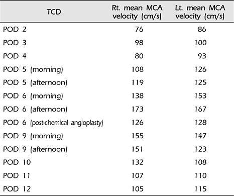
 | Fig. 1A 50-year-old woman reported at the emergency department with thunderclap headache and vomiting. (A) A non-enhanced brain computed tomography (CT) showed a subarachnoid hemorrhage in the suprasellar cistern, both sylvian and interhemispheric fissure. (B) Brain angiography CT revealed an aneurysm of the anterior communicating artery. The aneurysm was left dominant, inferior in direction, and multi-lobulated.
|
 | Fig. 2A transfemoral cerebral angiography was performed pre operatively. (A, B) TThe recurrent artery of Heubner (RAH) (arrows) originated bilaterally from the junction of the anterior cerebral artery and the anterior communicating artery. The mean diameter of right and left RAH was 8 mm and 4 mm, respectively.
|
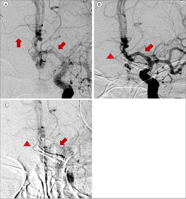 | Fig. 3(A) Both the recurrent artery of Heubner (RAH) (arrows) were evidently visible on the pre-operative transfemoral cerebral angiography (TFCA). (B) On the TFCA performed 6 days postoperatively before chemical angioplasty, a blurred right RAH (arrowhead) was seen due to decreased perfusion. The left RAH (arrow) was not visible. (C) On the TFCA performed after chemical angioplasty, the perfusion of right RAH (arrowhead) improved and left RAH (arrow) was visible.
|
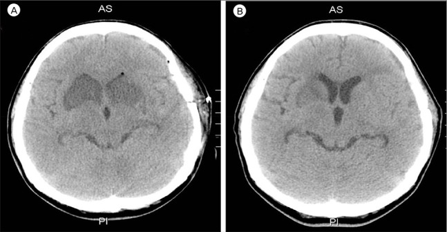 | Fig. 4(A) A 7-day postoperative follow-up brain computed tomography (CT) showed a newly developed low density lesion in the recurrent artery of Heubner (RAH) territory bilaterally, caudate nucleus, anterior portion of basal ganglia, and internal capsule. (B) The 14-day postoperative follow-up brain CT showed no progression of the low density lesion. The low density lesion in both of the RAH territory was blurred.
|
Table 1
Mean MCA velocity measured by TCD
![]()
Go to :
DISCUSSION
The RAH exhibited a mean outer diameter of 0.8 ± 0.04 mm (range, 0.2–1.5) and a mean length of 23.4 ± 1.1 mm (range, 12–38). The origin of the RAH occurs most commonly at the junction of the ACA and ACoA, then from the proximal A2 segment, and finally from the distal A1 segment.4)7)9) The RAH supplies parts of the caudate nucleus, basal ganglia, and anterior limb of the internal capsule.9)
There are only a few papers in the literature that specifically describe the territory affected by RAH infarction.1)3) RAH occlusion can manifest as various symptoms, including weakness of the contralateral face and arm without sensory loss, transient aphasia, dysarthria, cognitive and behavioral abnormalities, abulia, agitation, hyperactivity, contralateral neglect, obsessive-compulsive disorder, and memory dysfunction (i.e., poor ability to perform declarative and procedural memory tasks). However, most RAH infarctions are accompanied by only transient neurological deficits, if any.1)2)10)11)12) The possible causes of cerebral infarction following SAH are cerebral vasospasm, microthrombo-embolism, cortical spreading ischemia, spasm in the microcirculation and impaired cerebral autoregulation. Operative maneuvers that can lead to blood flow disturbance and consequent infarction include inappropriate aneurysmal neck clipping, temporary occlusion of the parent artery, direct injury, retraction with a spatula, or trapping of the parent artery.12) In this case, multiple factors are considered to have caused infarction of the bilateral RAH territory, including the SAH corresponding to a Fisher Grade III, placement five times of temporary clips on the A1 segment, hypotension during surgery (systolic blood pressure less than 100 mmHg), and a mean arterial pressure of 65 mmHg. Cerebral vasospasm was also a significant causative factor in this case. The mechanism of vasospasm remains poorly understood. However, in the last 3 decades, certain mechanisms have been identified that might play a role in its development. Factors such as calcium-dependent and -independent vasoconstriction, breakdown of products in the blood of subarachnoid space, and imbalance between vasoconstrictor and vasodilator substances derived from the endothelium may be major causes of vasospasm.8) Cerebral vasospasm post-SAH may involve the complete arterial system from the circle of Willis up to the distal vessel segments.15) A high volume of cisternal hemorrhage on an admission CT scan in a patient with Fisher Grade III aneurysmal SAH was found to be highly associated with delayed ischemic neurological deficit (DIND) due to cerebral vasospasm.6) When the subarachnoid blood went undetected or was distributed diffusely, severe vasospasm was almost never encountered; however, in the presence of a large amount of SAH, severe vasospasm almost always occurred.5) Approximately 11–20% of episodes of DIND are characterized by cerebral infarction.13)14) Cerebral infarction mostly occurs in patients who develop angiographic vasospasm.14) Our patient was alert during her stay in the neuro-intensive care unit. As the mean MCA velocity increased, the patient became lethargic; this was attributed to DIND. Angiographic vasospasm was found in the preoperative and postoperative TFCA. Thus it was deduced that the decreased perfusion of both RAH due to cerebral vasospasm was a major cause of the bilateral RAH territorial infarction. In addition to cerebral vasospasm, the other causes of bilateral RAH infarction were assumed to be the repeated temporary clipping on the Lt. A1 segment and the presence of hypotension during surgery. The prolonged temporary clipping time of 26 minutes, 45 seconds and hypotension (systolic blood pressure less than 100 mmHg for 115 minutes and mean arterial pressure of 65 mmHg for 70 minutes) during surgery likely significantly lowered cerebral blood perfusion and cerebral perfusion pressure. These conditions would have been sufficient to cause bilateral RAH infarction. During the temporary clipping, the systolic blood pressure was always below 100 mmHg; the mean arterial blood pressure was below 65 mmHg for half of the clipping time. Temporary clipping of the main cerebral artery can lead to infarcts of the perforating artery, and temporary clipping of the A1 segment can lead to infarction of the RAH.12) Sasaki et al.12) reported nine patients of RAH infarction following aneurysm clipping, all of whom had ACoA aneurysm, in a population of 1,045 patients with treated aneurysms and 238 with ACoA aneurysms. The incidence of the causes of RAH infarctions: A1 temporary occlusion (four), retraction (three) and injury of RAH (two). In this case, vasospasm was considered to be a more likely cause of RAH infarction than temporary occlusion or hypotension. This inference was made based on the fact that immediately following the operation the patient had no neurologic deficit or abnormal mental status. The patient's reduced mental status appeared alongside the increase in the mean MCA velocity. There were no abnormal findings in the post-operative brain CT. An intra-operative Doppler was applied and intact flow of the RAH was observed.
Go to :
CONCLUSION
It is important to keep in mind that the clipping of an ACoA aneurysm can cause infarction of the RAH. In cases of RAH infarction following aneurysm surgery, the aneurysm is almost always in the ACoA.12) When a patient has DIND with an elevated mean MCA velocity after clipping of an ACoA aneurysm, neurosurgeons should consider the possibility of an RAH infarction caused by decreased perfusion due to cerebral vasospasm.
Go to :
 XML Download